


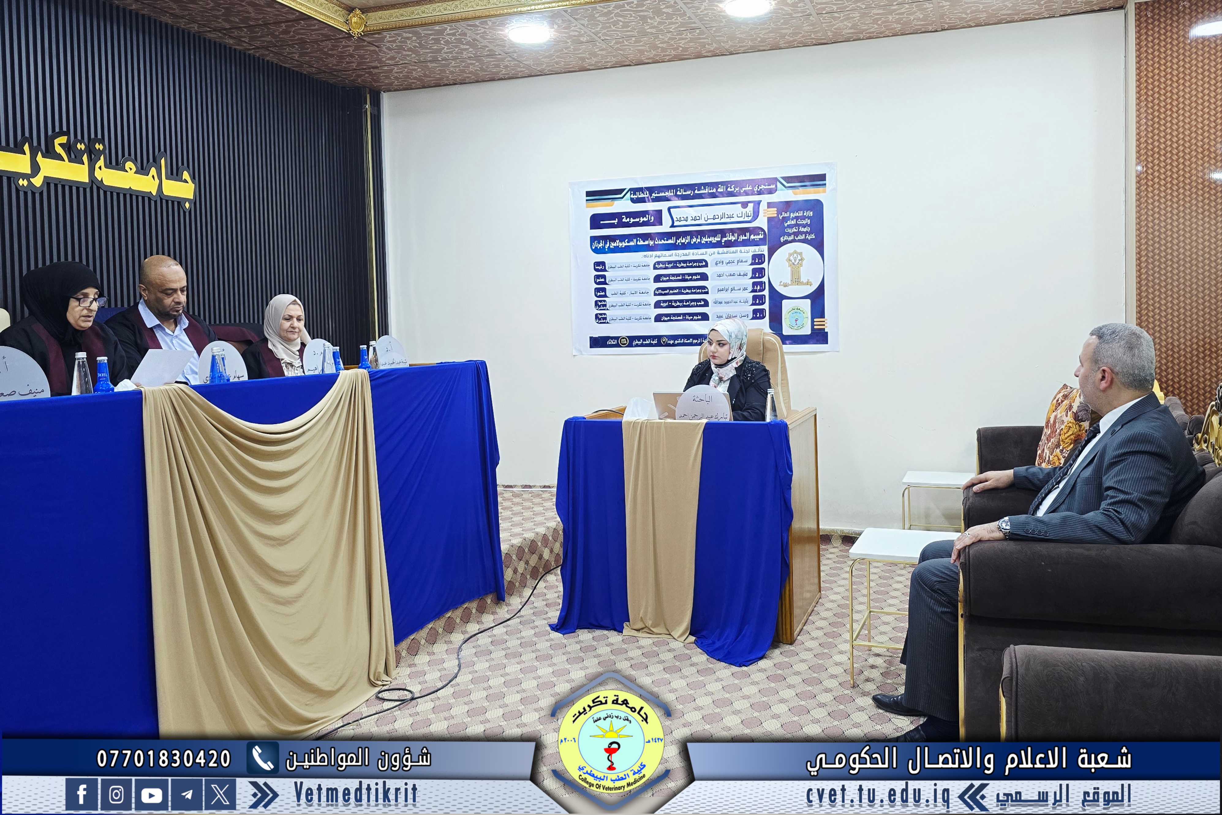
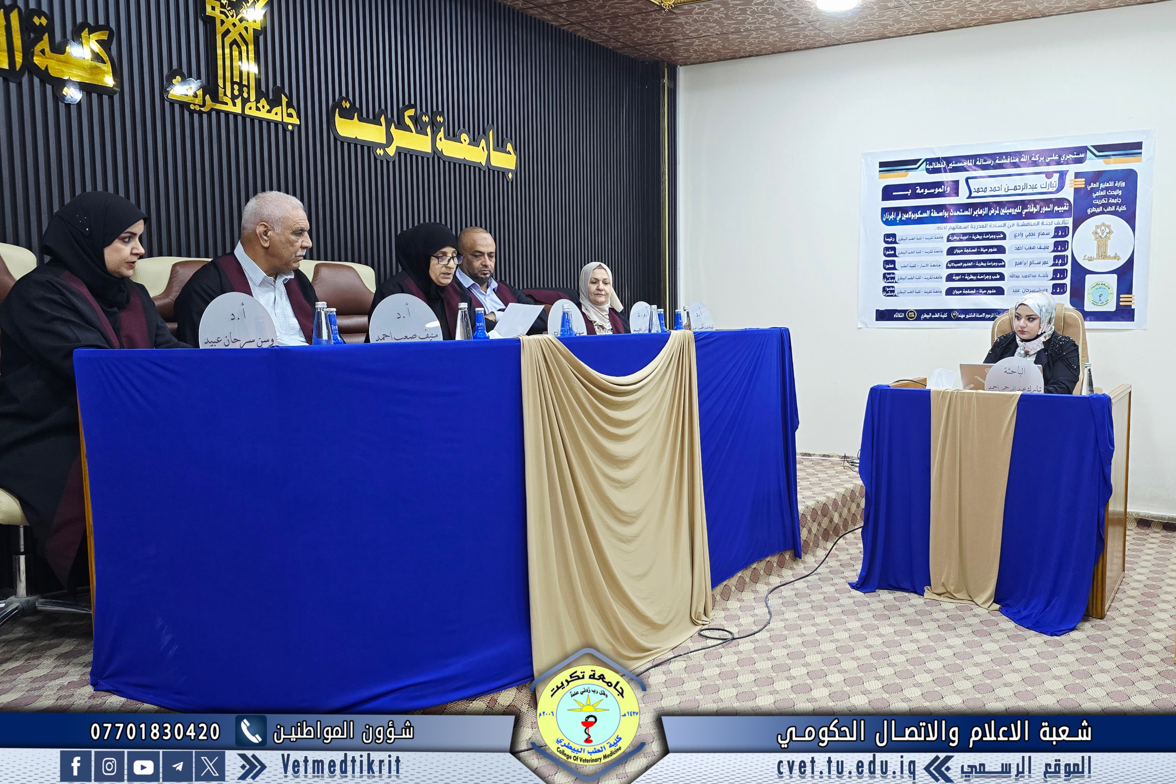
With the grace of Almighty God, a master’s thesis defense was held at the College of Veterinary Medicine, Tikrit University, attended by Professor Dr. Bashar Sadiq Noami, Dean of the College.
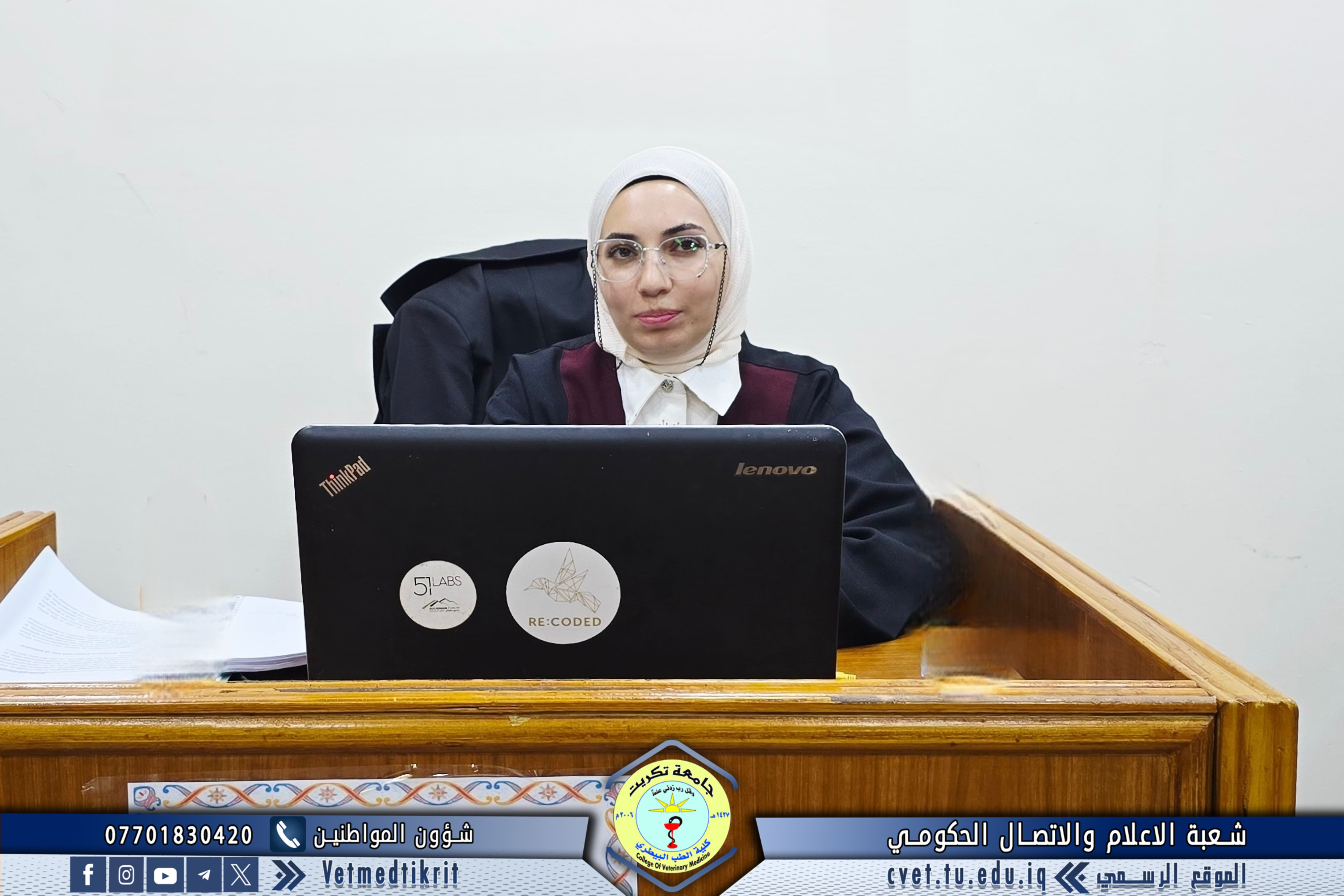
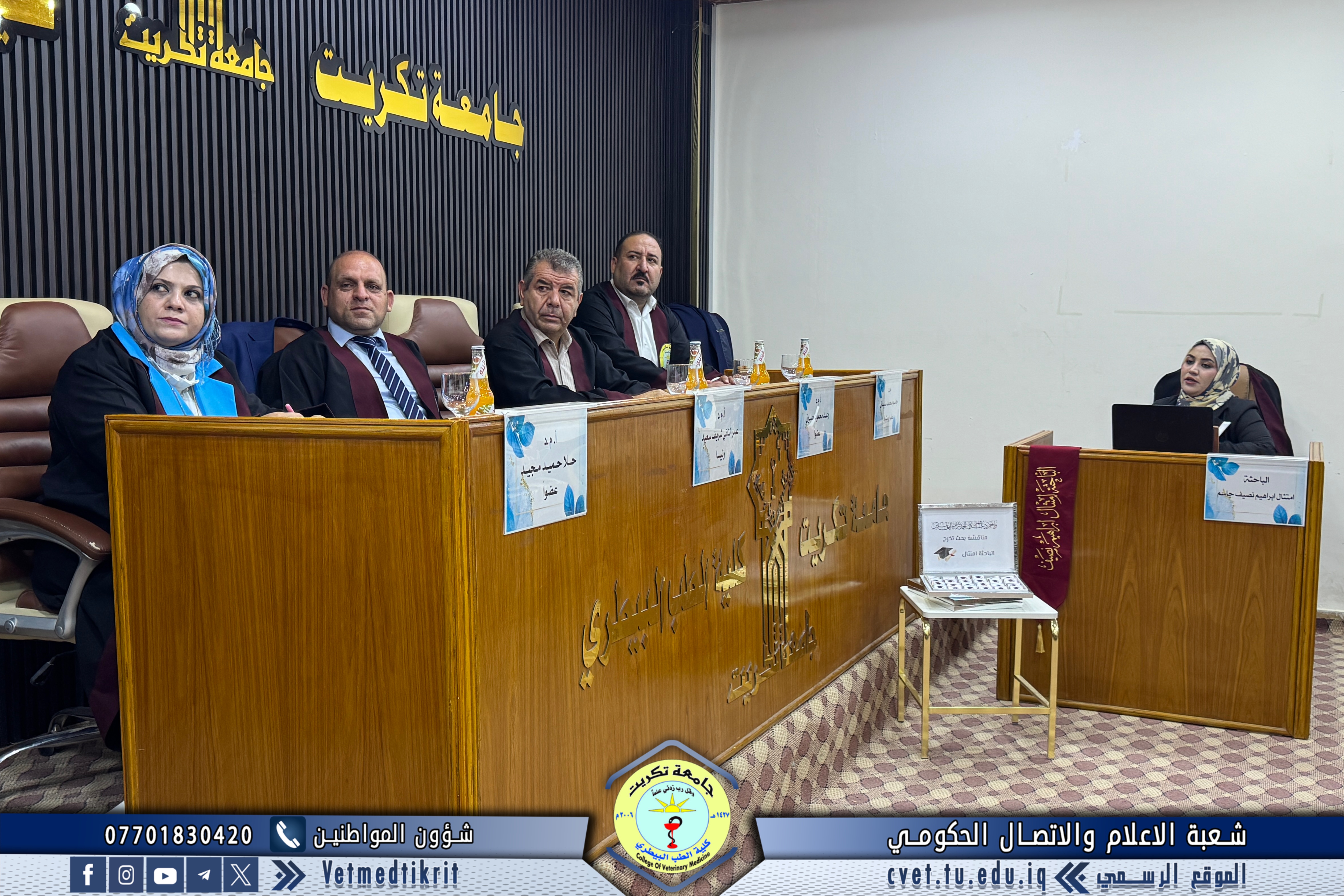
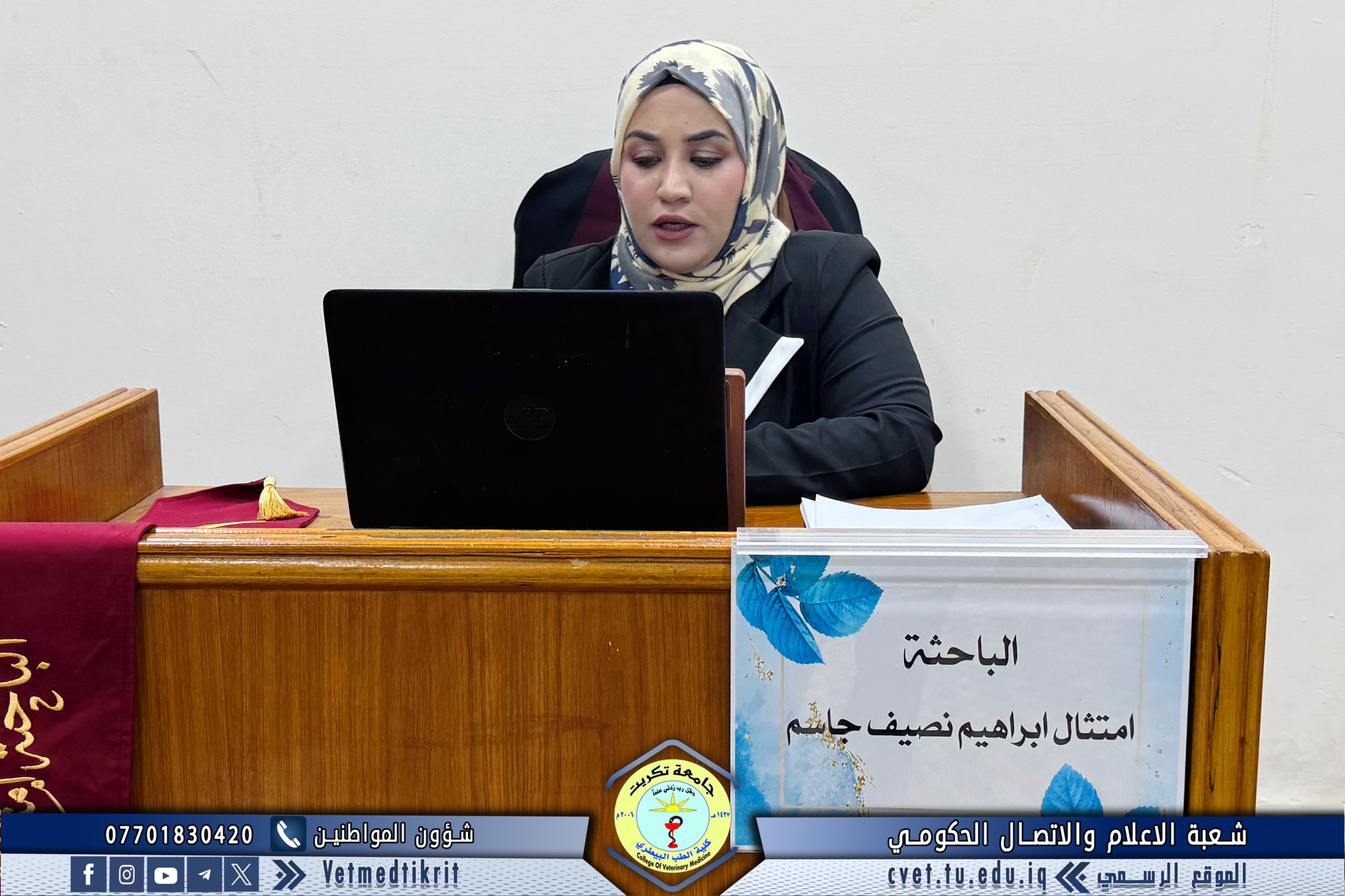
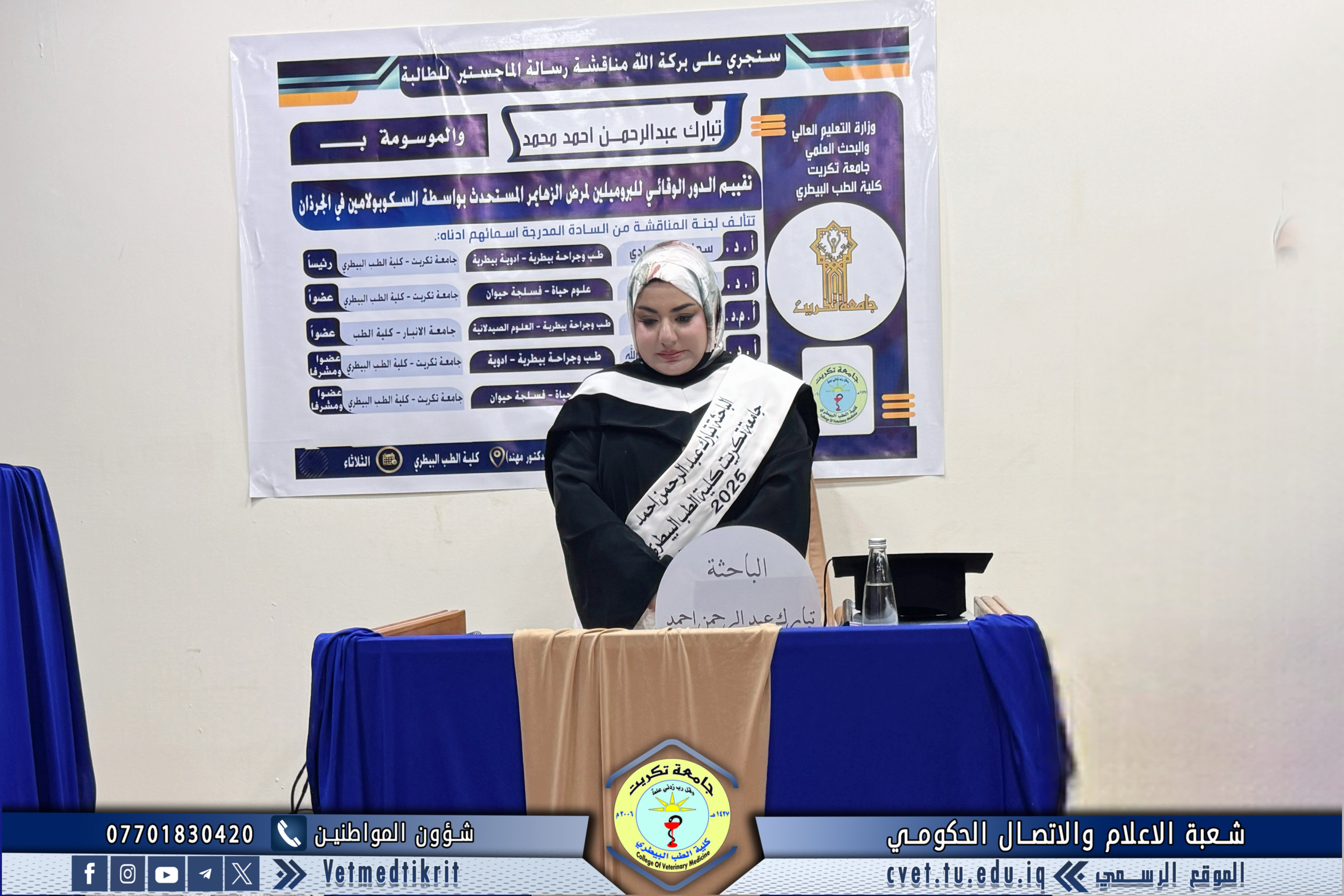
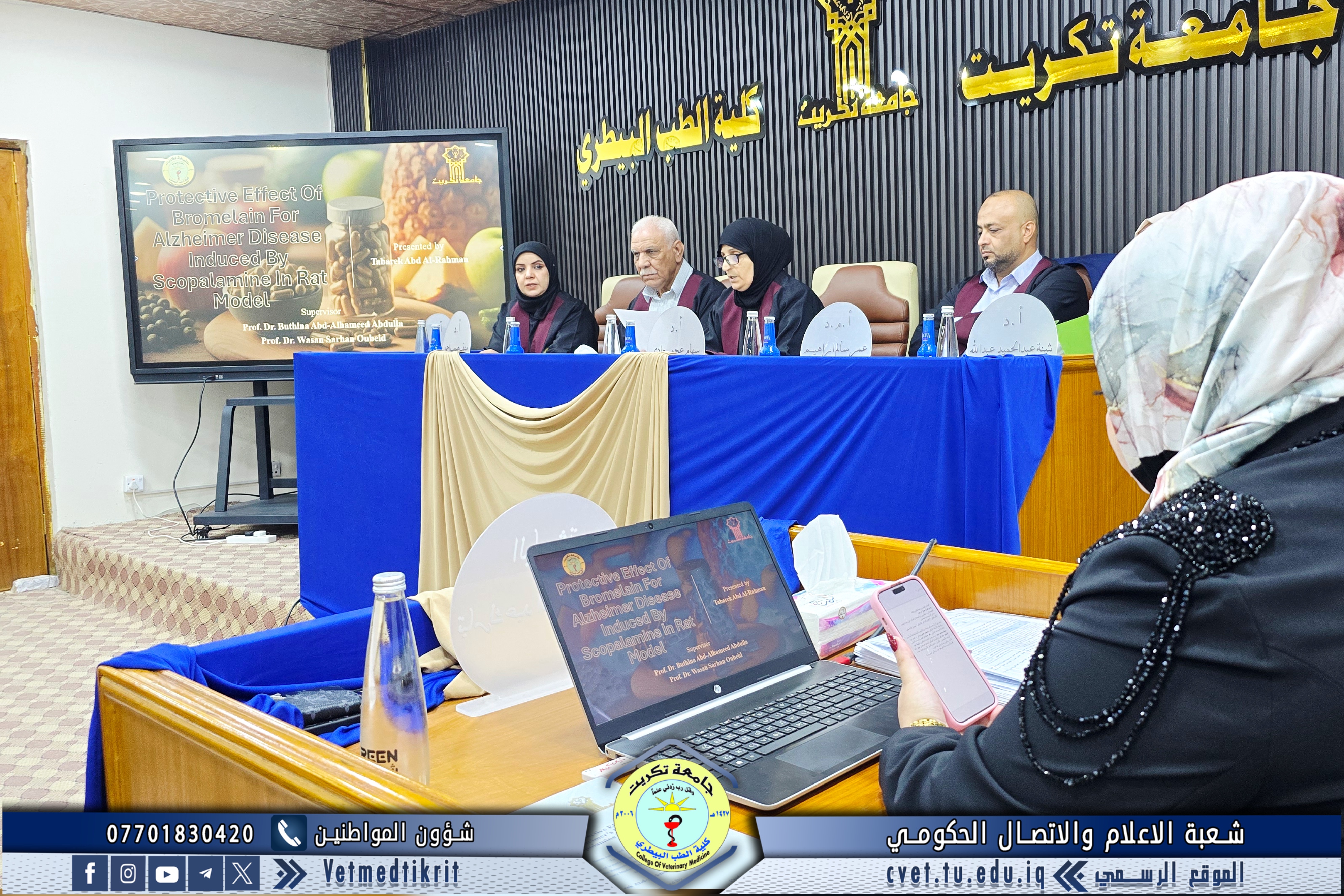
The thesis, titled “Evaluation of the Protective Role of Bromelain Against Scopolamine-Induced Alzheimer’s Disease in Rats”, was presented by graduate student Tabarak Abdulrahman Ahmed in the specialization of Veterinary Medical Pharmacology.
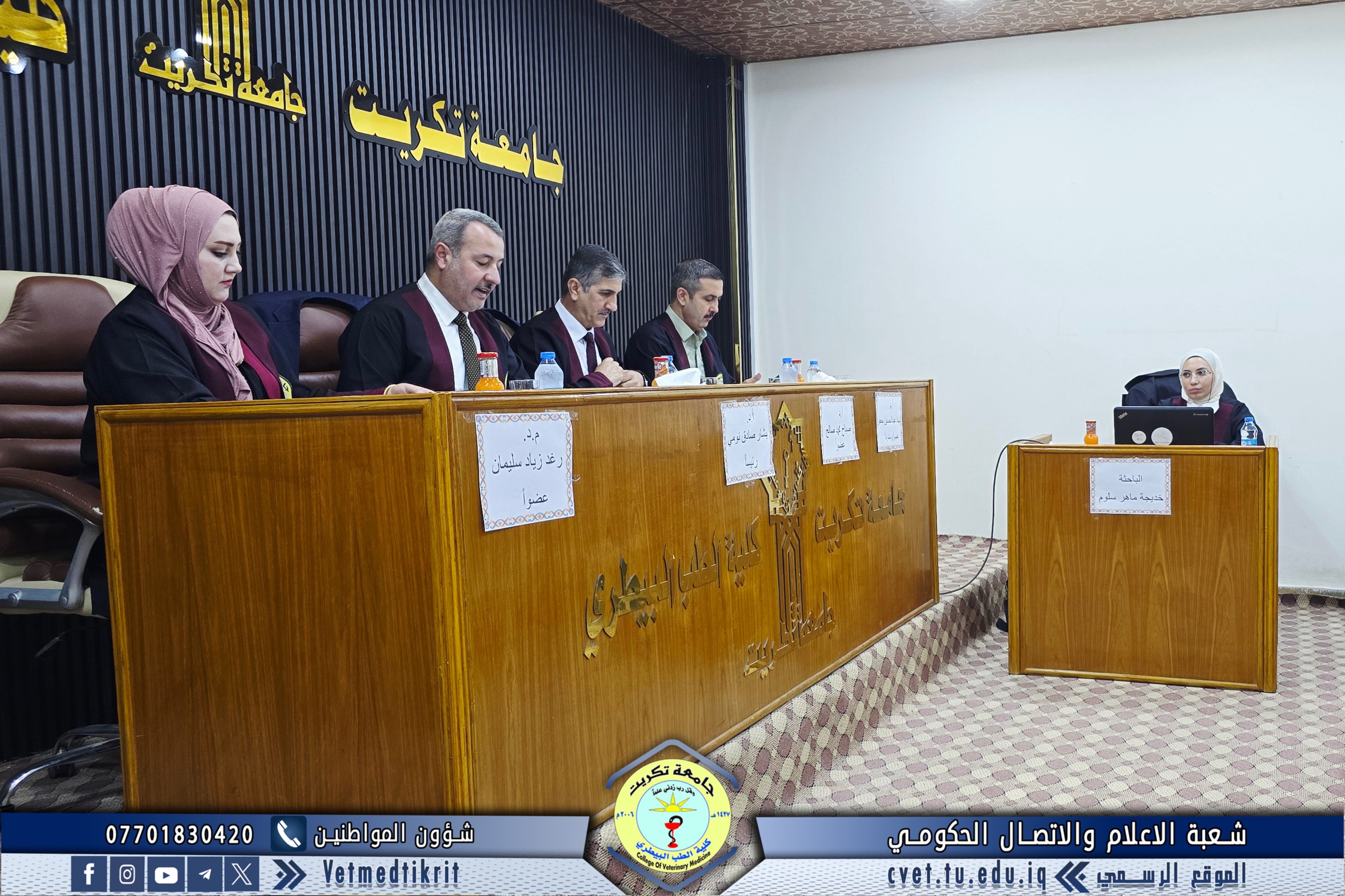
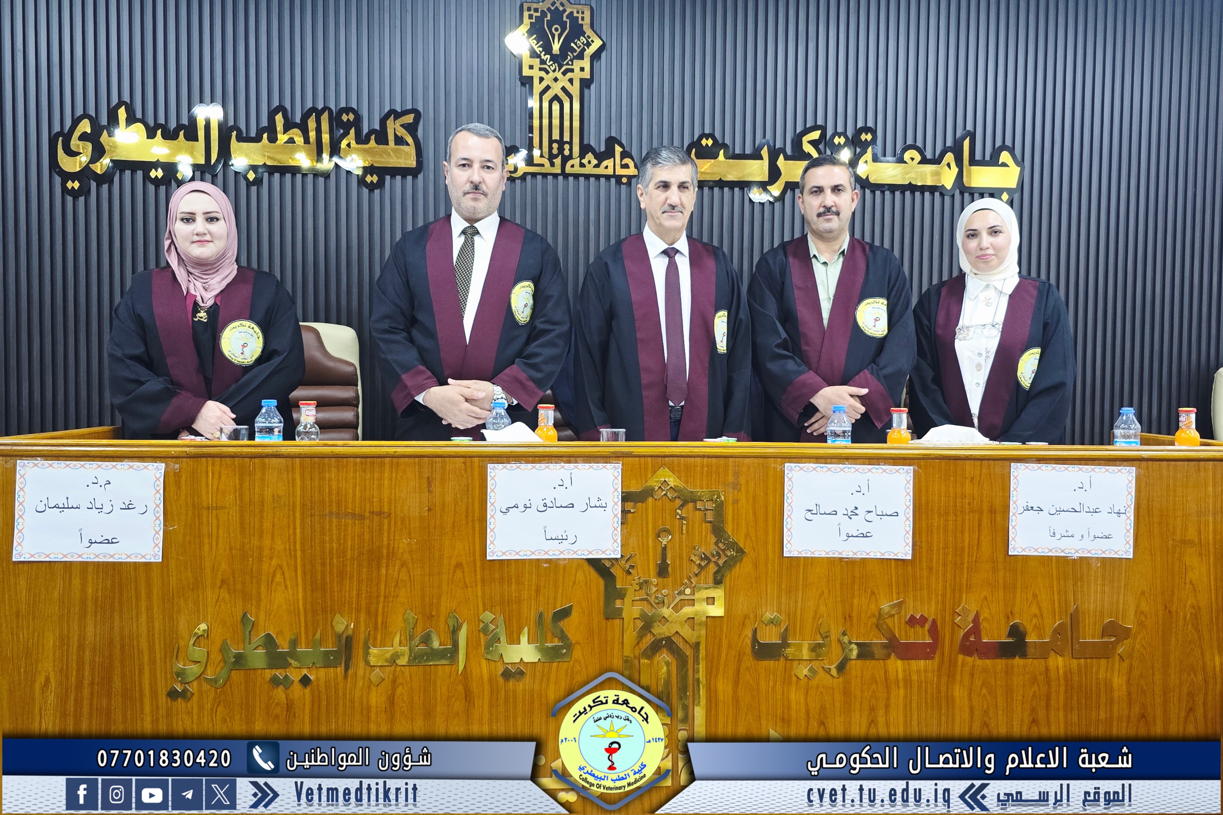
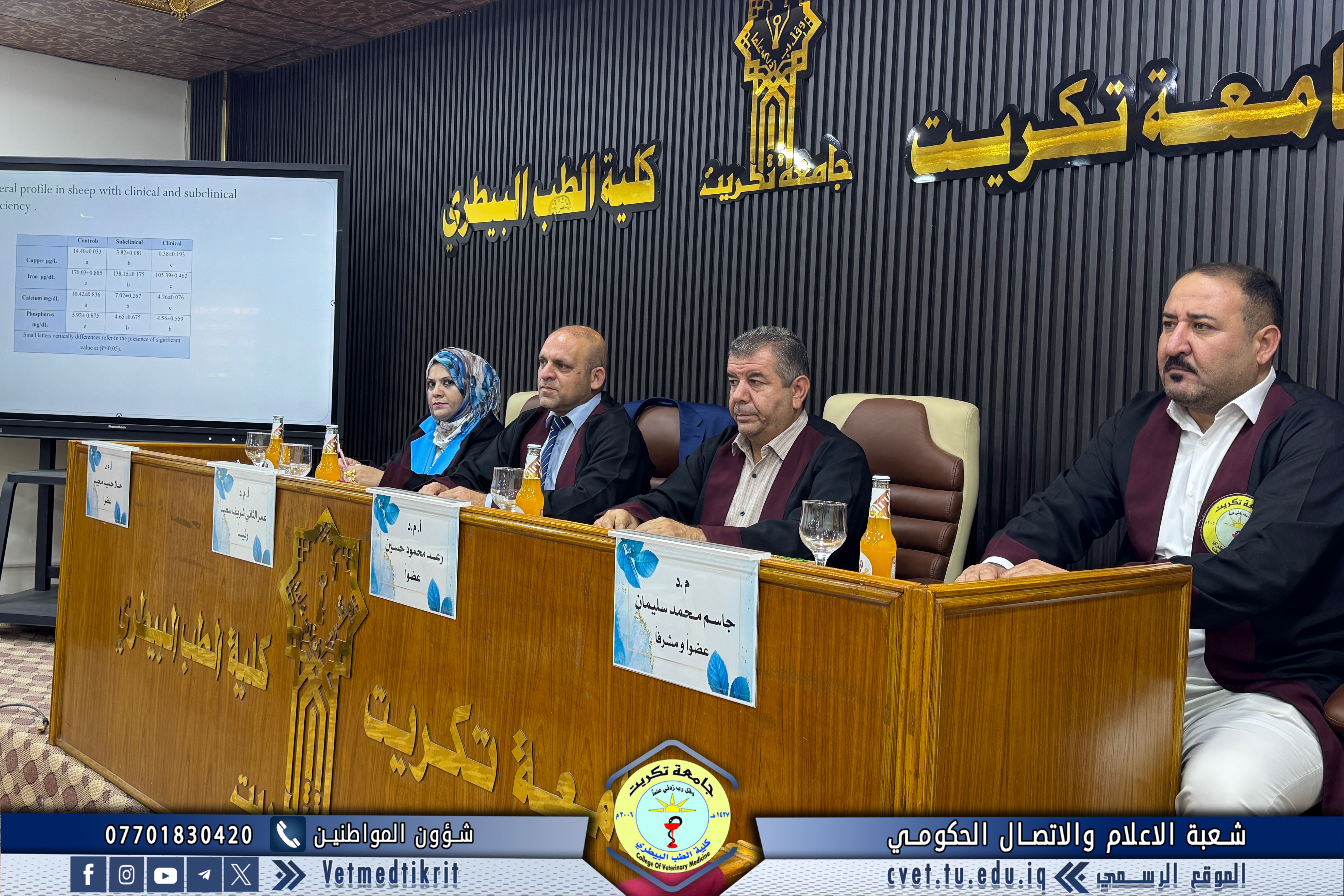
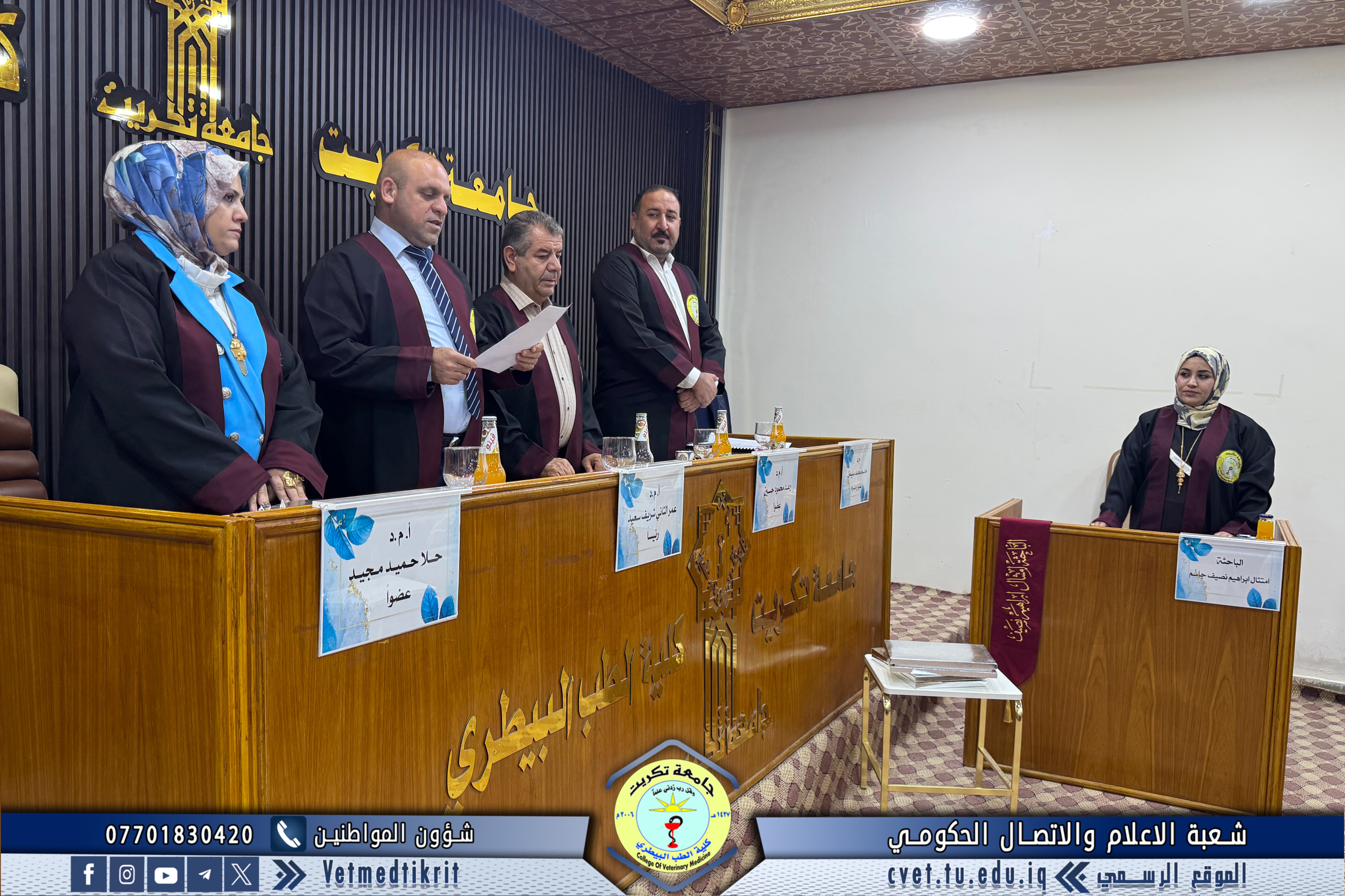
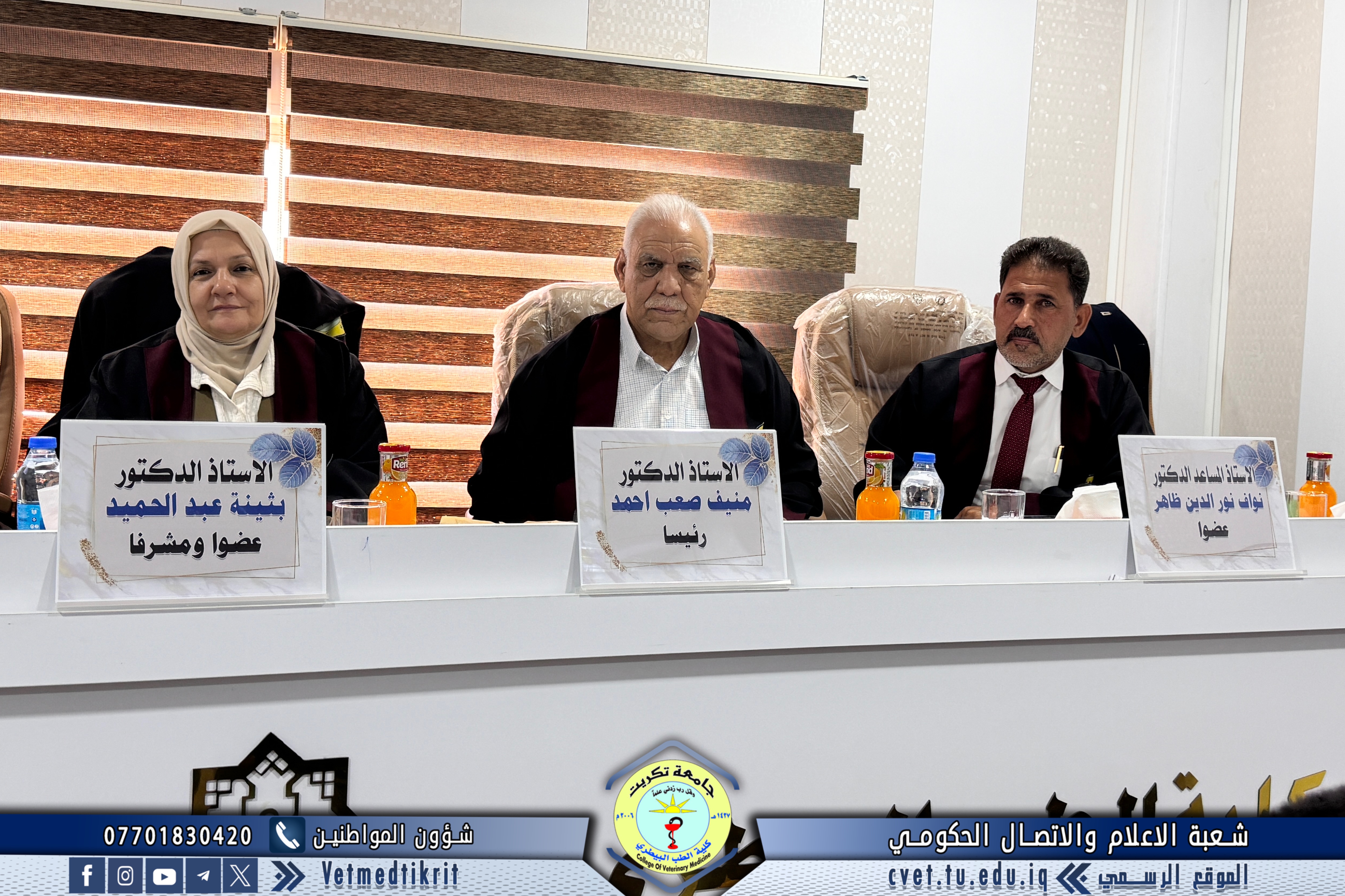
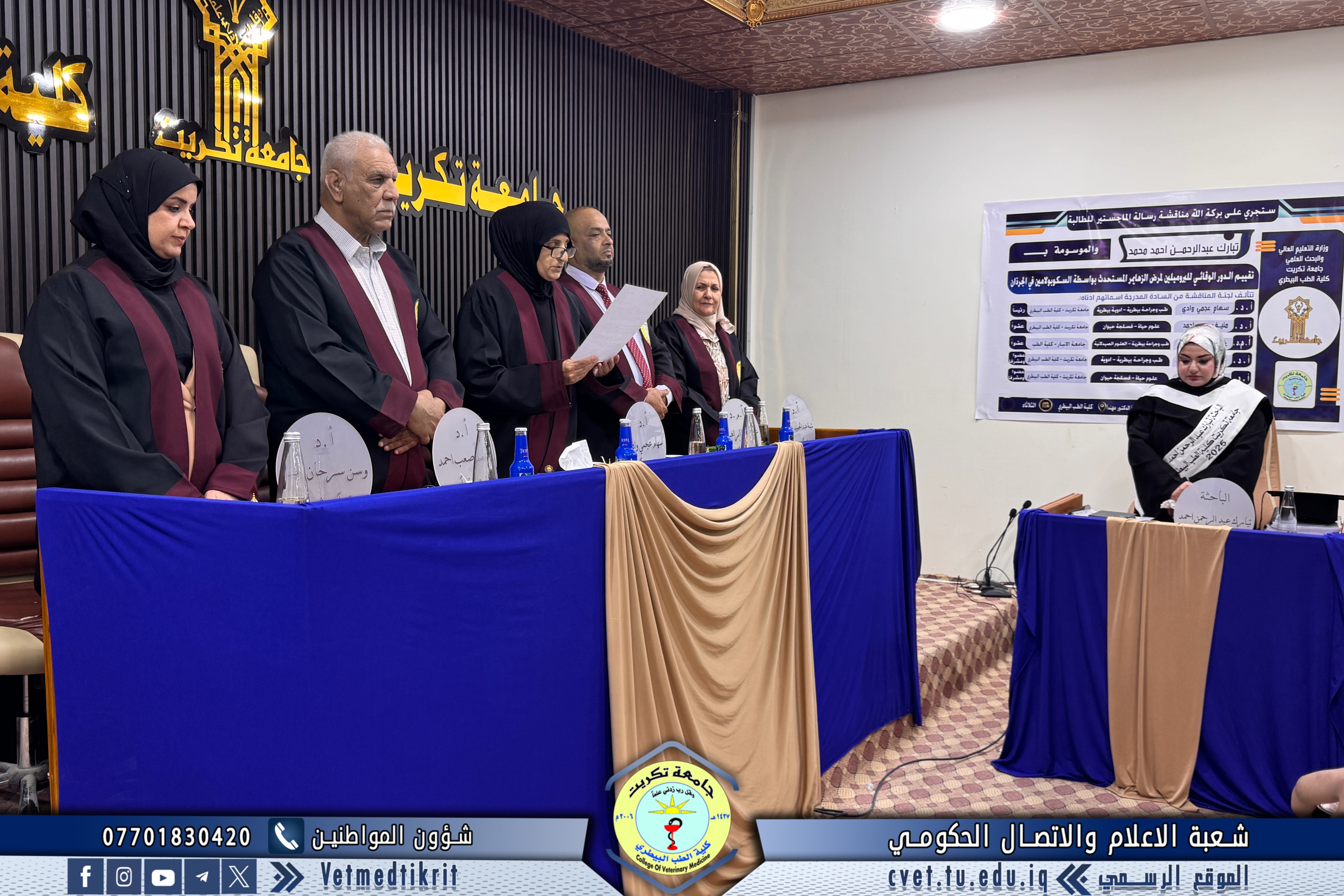
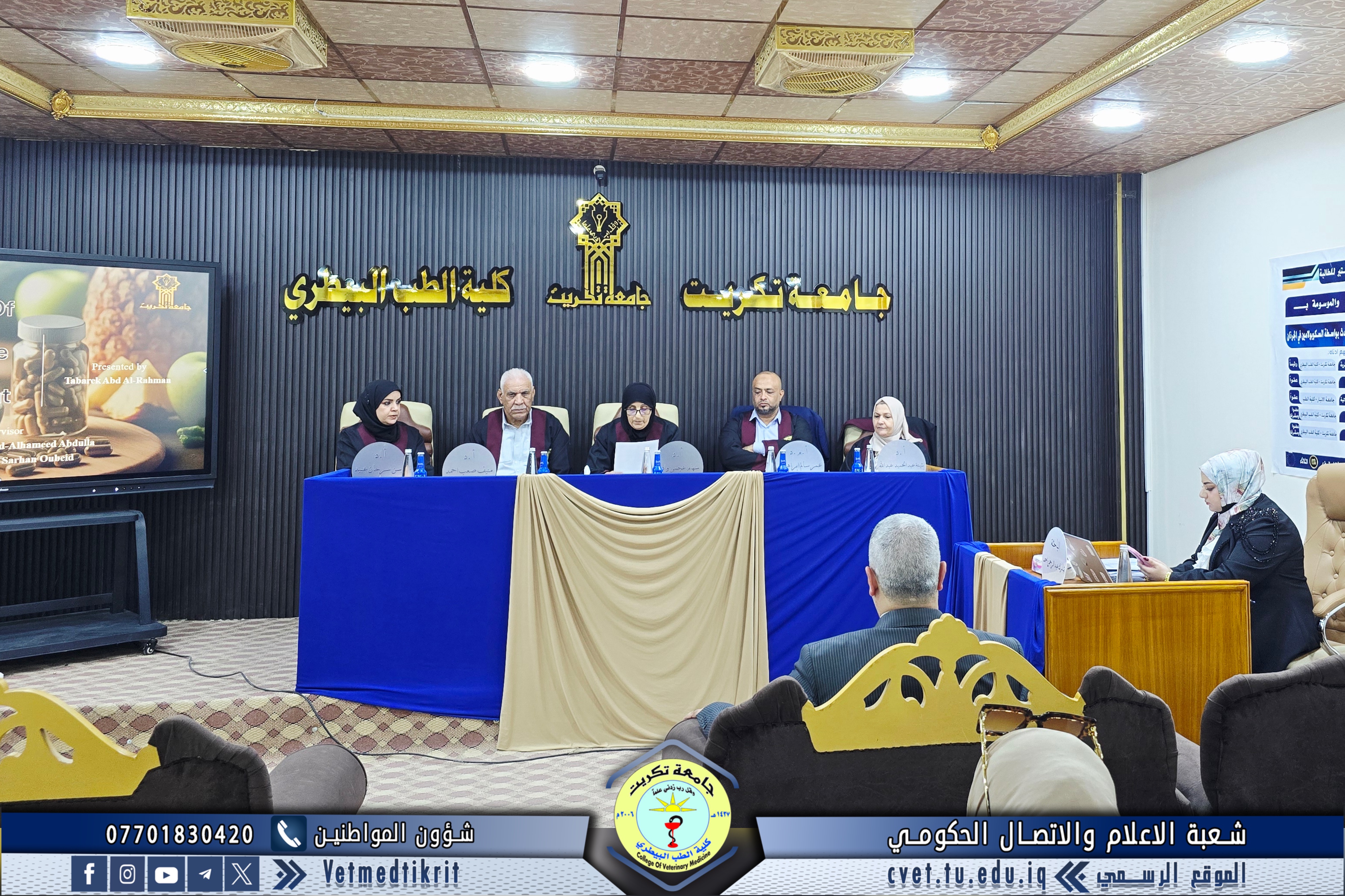
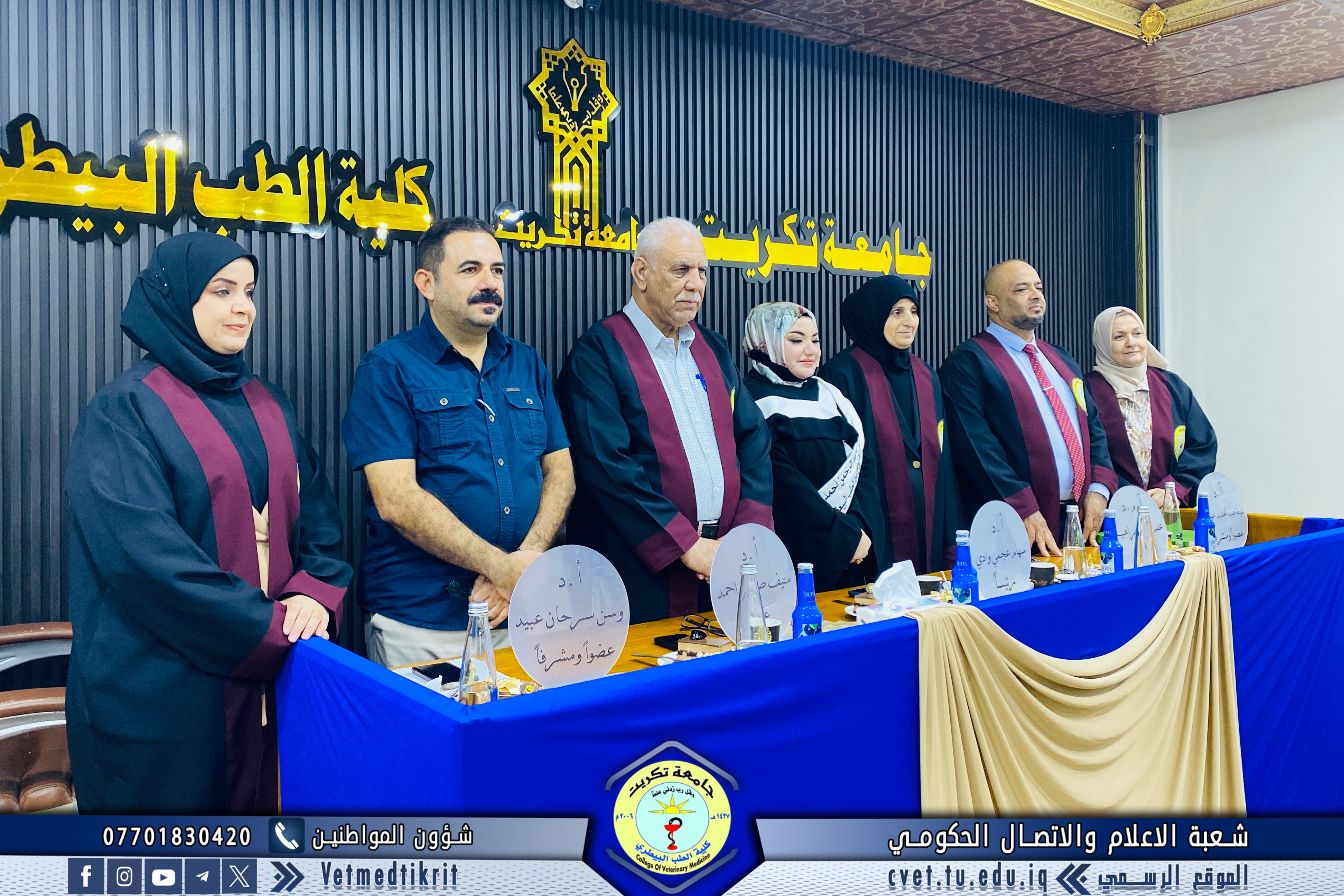
The examination committee consisted of the following members:
Professor Dr. Siham Ajami Wadi – Veterinary Pharmacology / Tikrit University, College of Veterinary Medicine – Chair
Professor Dr. Muneef Saeb Ahmed – Animal Physiology / Tikrit University, College of Veterinary Medicine – Member
Assistant Professor Dr. Omar Salem Ibrahim – Pharmaceutical Sciences / University of Anbar, College of Medicine – Member
Professor Dr. Buthaina Abdulhameed Abdullah – Pharmacology / Tikrit University, College of Veterinary Medicine – Member and Supervisor
Professor Dr. Wasan Sarhan Ubaid – Animal Physiology / Tikrit University, College of Veterinary Medicine – Member and Supervisor
Summary of the Thesis:
The present study aimed to evaluate the protective role of bromelain against scopolamine-induced Alzheimer’s disease through its antioxidant and anti-inflammatory activities, as well as to assess its effectiveness in inhibiting acetylcholinesterase (AChE) activity and enhancing acetylcholine (ACh) levels to improve memory function.
A total of 25 mature female rats, aged 10–12 weeks and weighing an average of 207 g (ranging from 181–204 g), were used. The experiment was conducted from August 29, 2024, to September 11, 2024, at the animal house of the College of Veterinary Medicine, Tikrit University.
The animals were divided into five main groups (five rats each):
Negative control group
Positive control group
Bromelain → Scopolamine + Donepezil group
Bromelain → Scopolamine + Bromelain group
Pre-treated Bromelain group – received bromelain for 7 days, then scopolamine (0.02 mg/kg) + a combination of bromelain and donepezil (5 mg/kg) from day 8 to day 14.
Biochemical Findings:
The study measured levels of AChE, ACh, Aβ1–42, GSH, IL-1, and BCS among the groups.
AChE levels showed a significant increase (P ≤ 0.05) in the positive control group (506.5 ± 51.7) compared to the control (338.0 ± 38.2). Treated groups showed elevated AChE levels compared to control but lower than the positive group.
ACh levels decreased significantly (122.4 ± 23.1) in the positive group (P ≤ 0.05) compared to the control (181.2 ± 28.2). Treated groups showed no significant difference compared to control.
Aβ1–42 levels increased significantly (1461.2 ± 130.4) in the positive group (P ≤ 0.05) compared to the control (1230.0 ± 124.2), while treated groups showed a significant decrease.
GSH levels significantly decreased (21.408 ± 0.399) in the positive group (P ≤ 0.05) compared to the control (26.056 ± 0.711), while treated groups showed a significant increase.
IL-1 levels increased significantly (156.8 ± 26.4) in the positive group compared to the control (128.6 ± 23.6), while treated groups showed a significant reduction.
BCS levels increased significantly (121.200 ± 25.20) in the positive group (P ≤ 0.05) compared to control (112.020 ± 20.09), while treated groups showed a marked decrease.
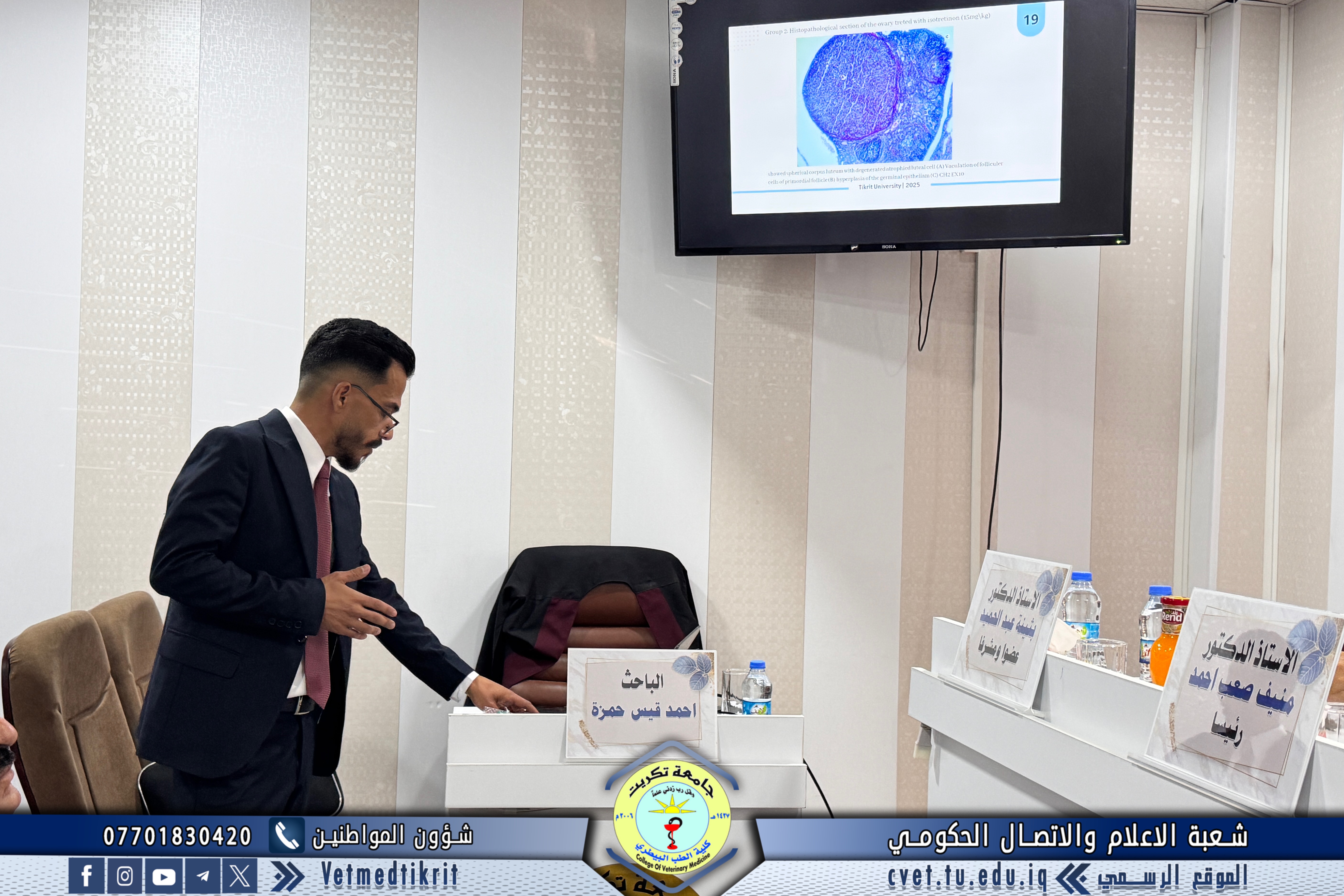
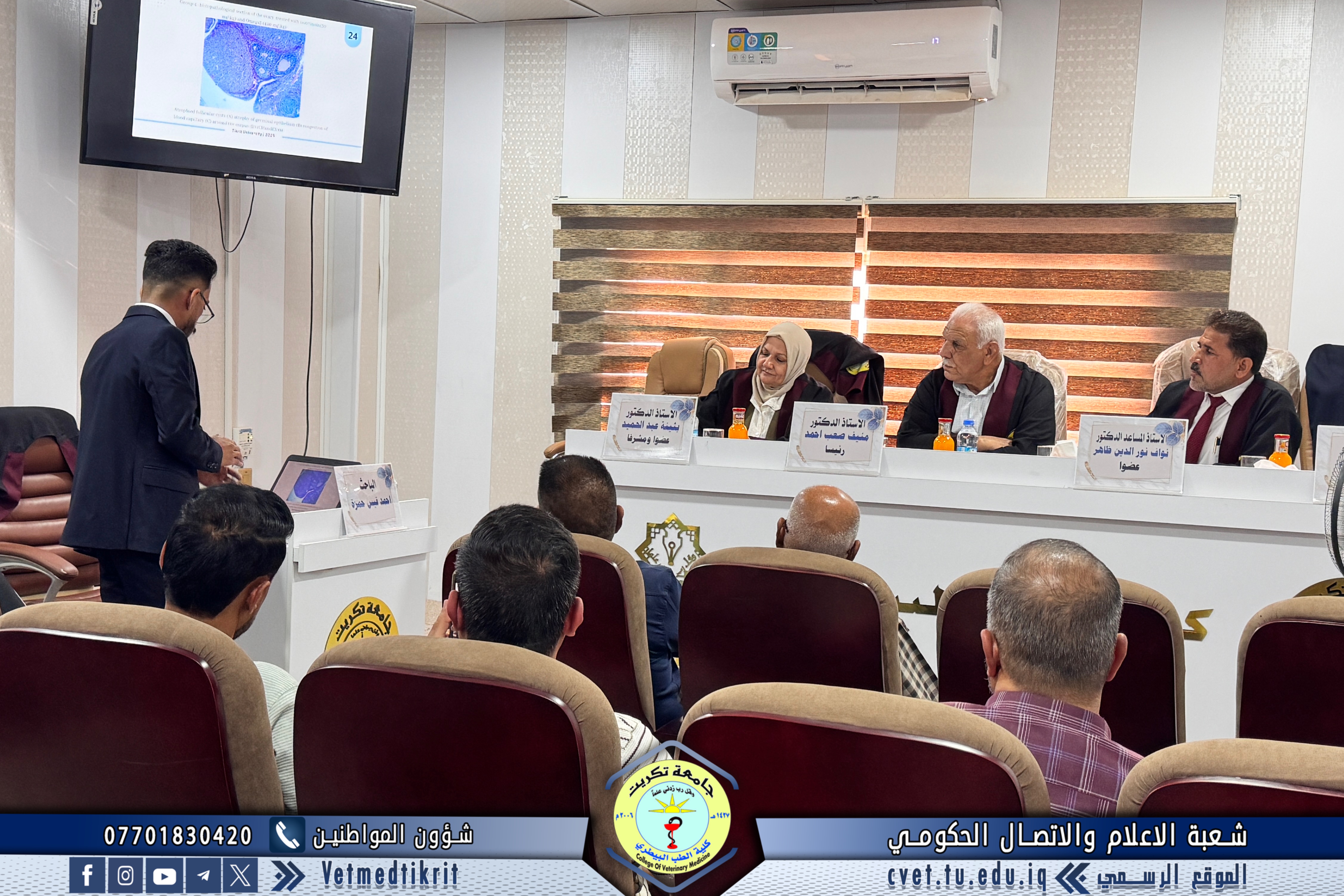
Histological Findings:
Histological examination of the brain cortex in the scopolamine group revealed large vacuoles and scattered inner pyramidal cells surrounded by perinuclear vacuolar degeneration. Microvessels were surrounded by leukocytes.
In the third group, the internal pyramidal layer contained medium and small neurons with few glial cells, and microvessels were surrounded by neural bundles.
In the fourth group, microscopy showed the brain cortex covered with meningeal membranes containing meningeal vessels; the molecular layer consisted of a few small pyramidal cells surrounded by fine neural bundles, and the granular layer contained small and medium-sized pyramidal cells.
In the fifth group, the cortex was covered by a pyramidal meningeal membrane with organized molecular, granular, and pyramidal layers showing normal structure surrounded by neural fibers.
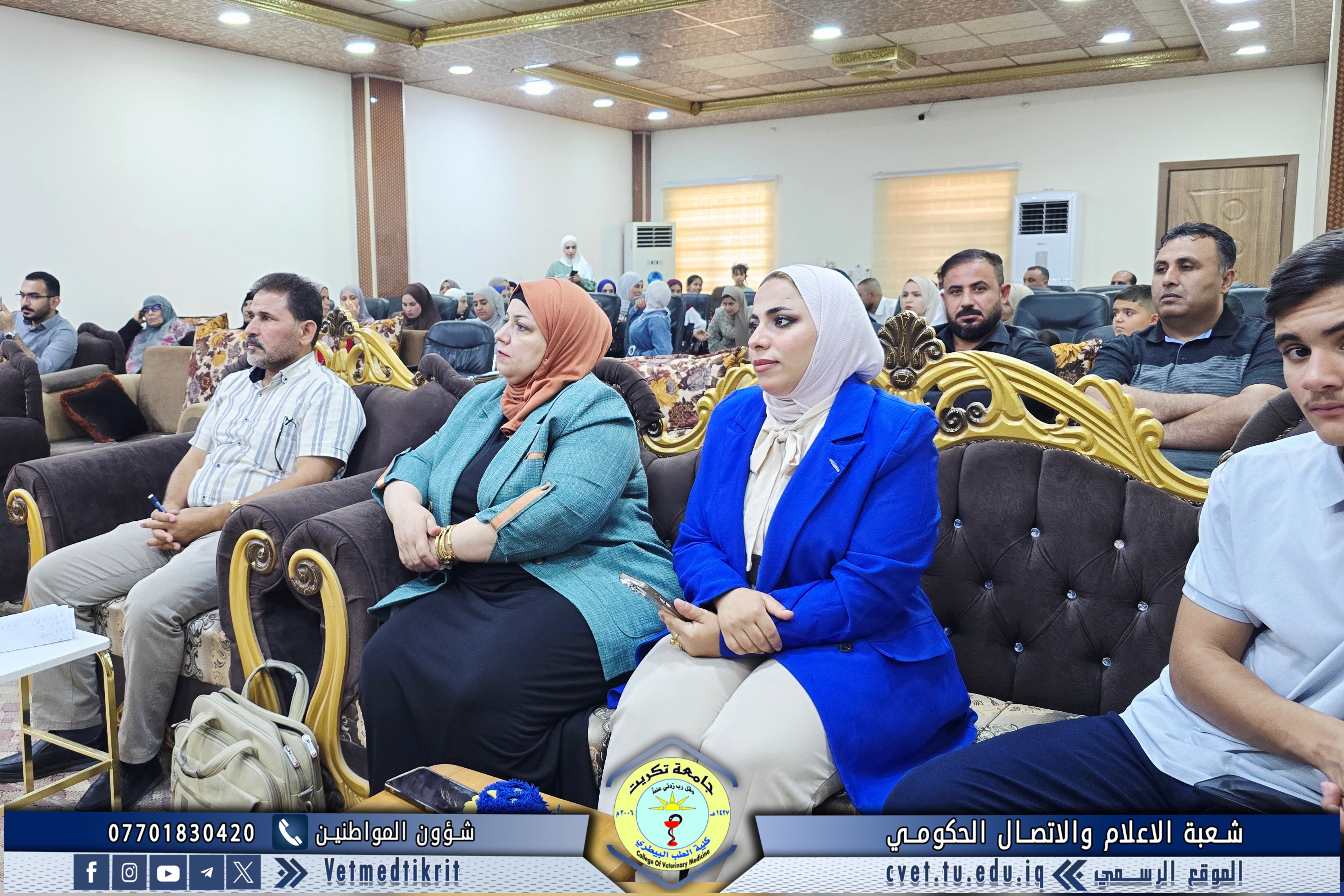
The defense session, held in Dr. Muhannad Maher Hall at the College of Veterinary Medicine, was attended by a number of faculty members and students.