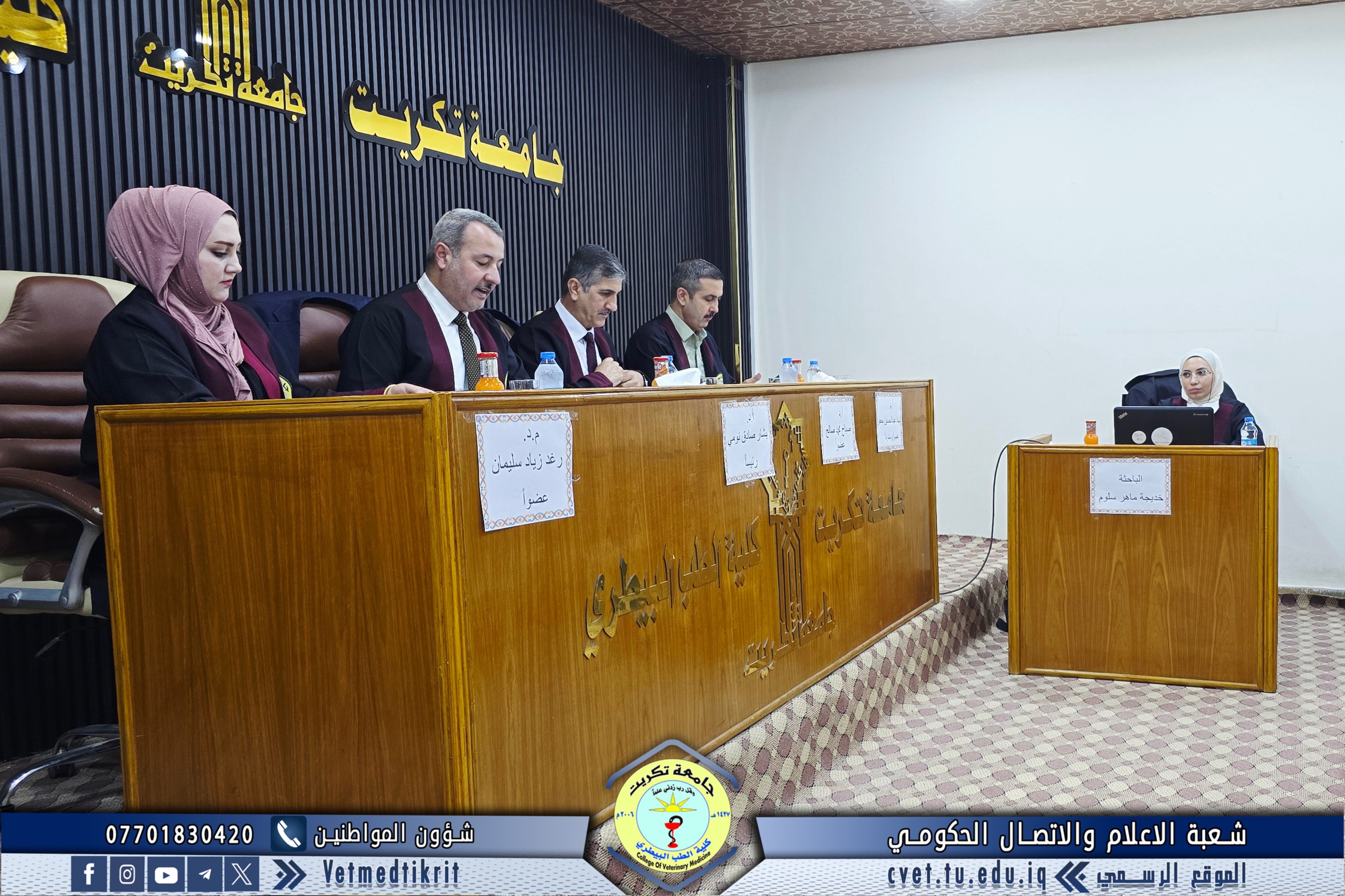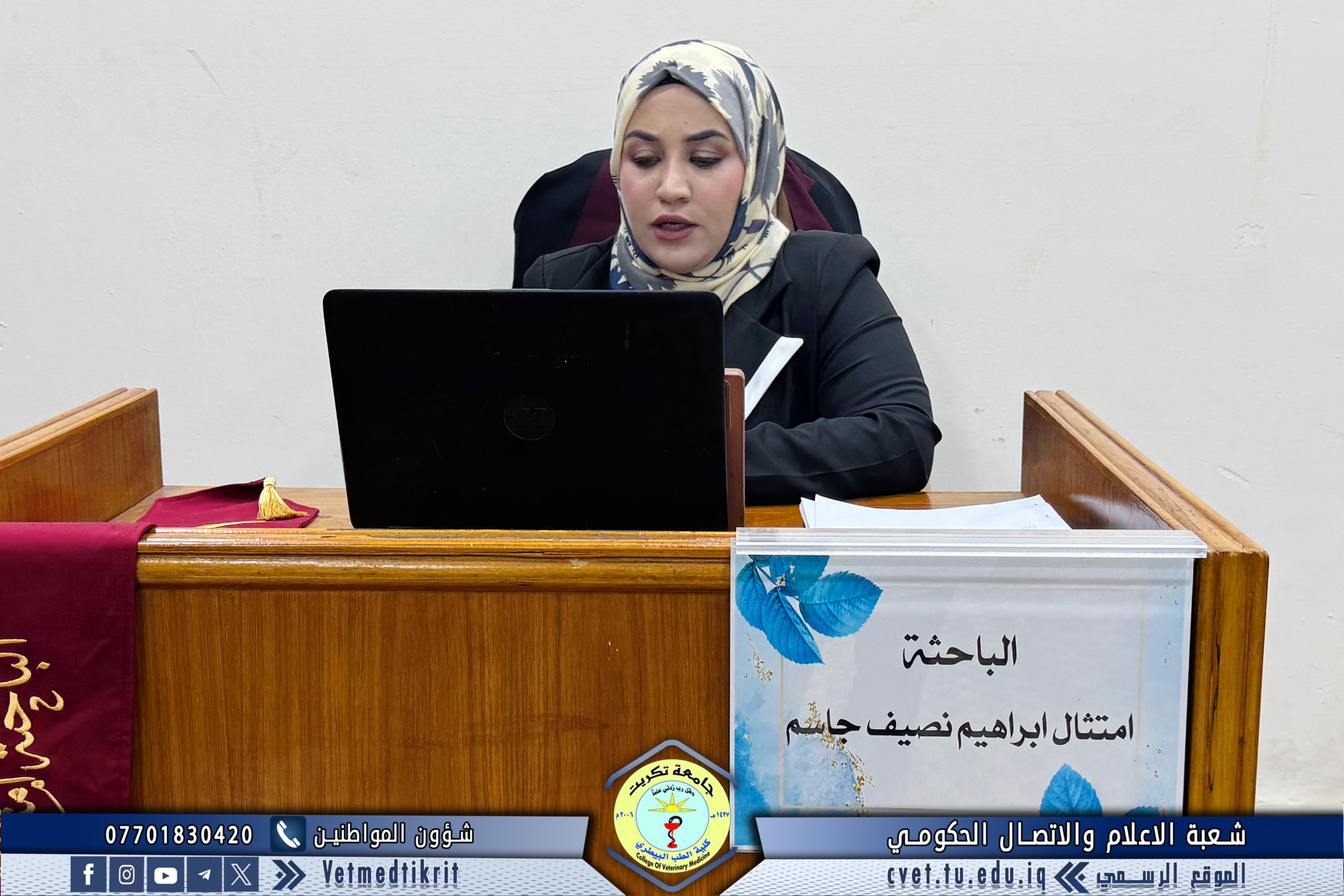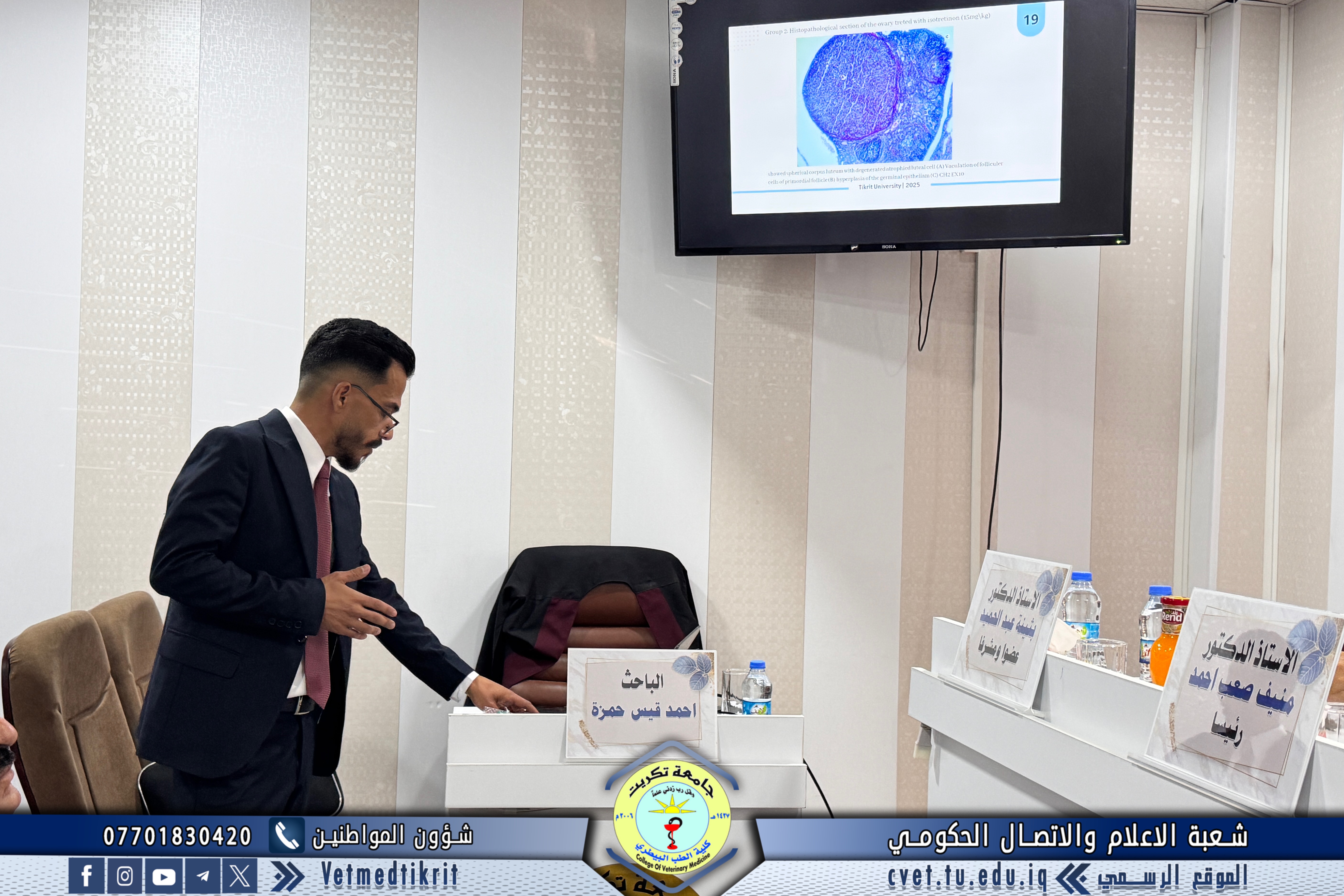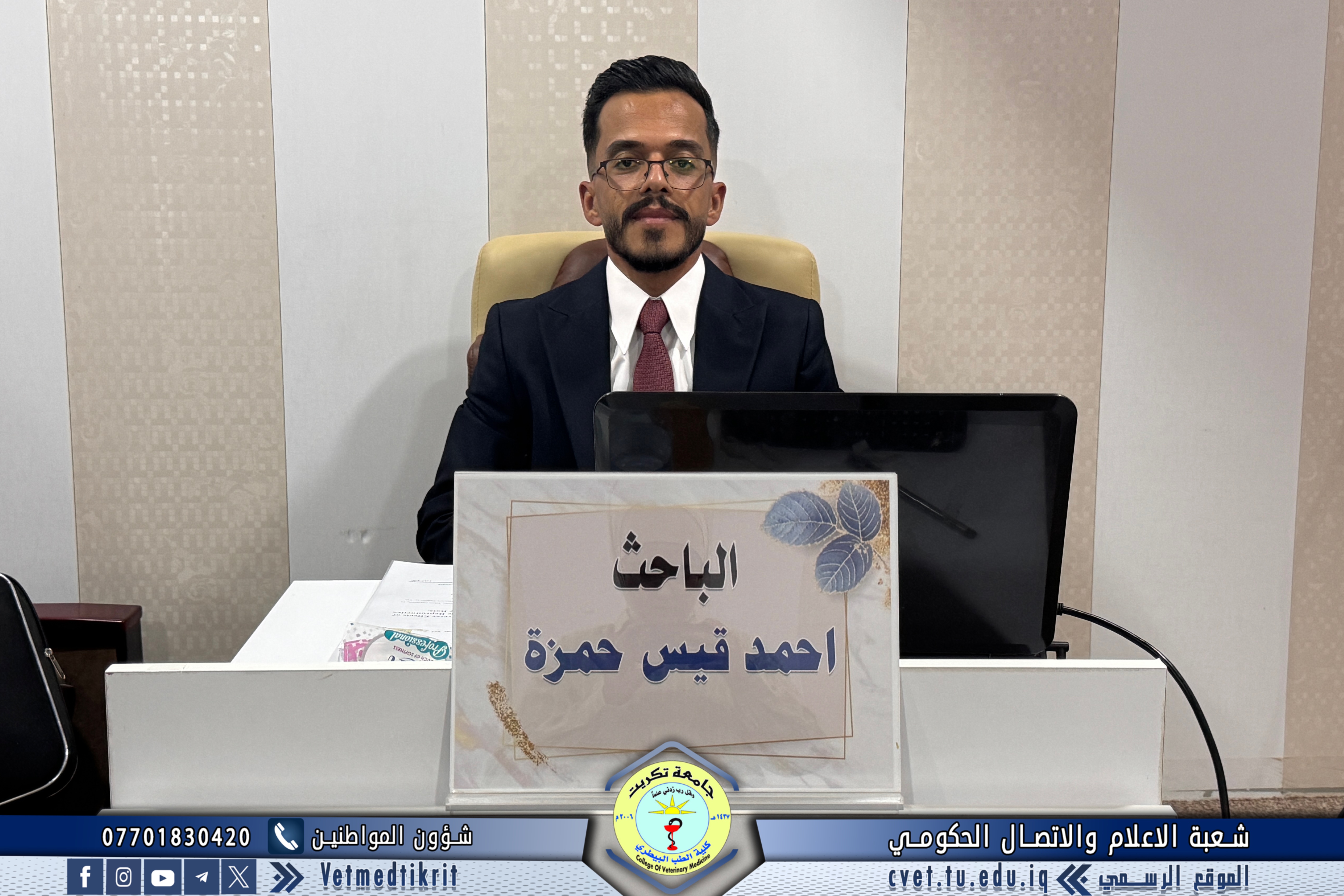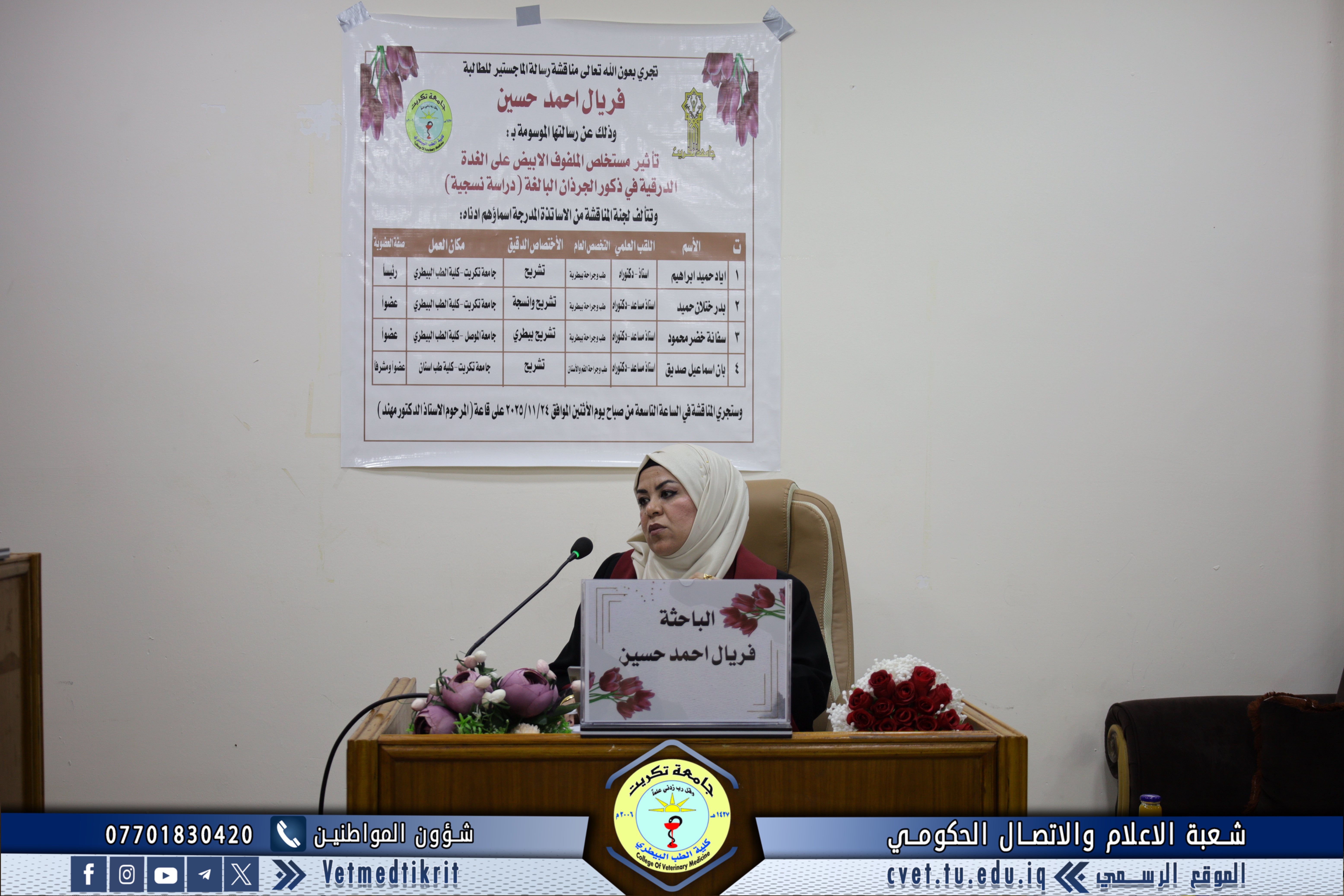
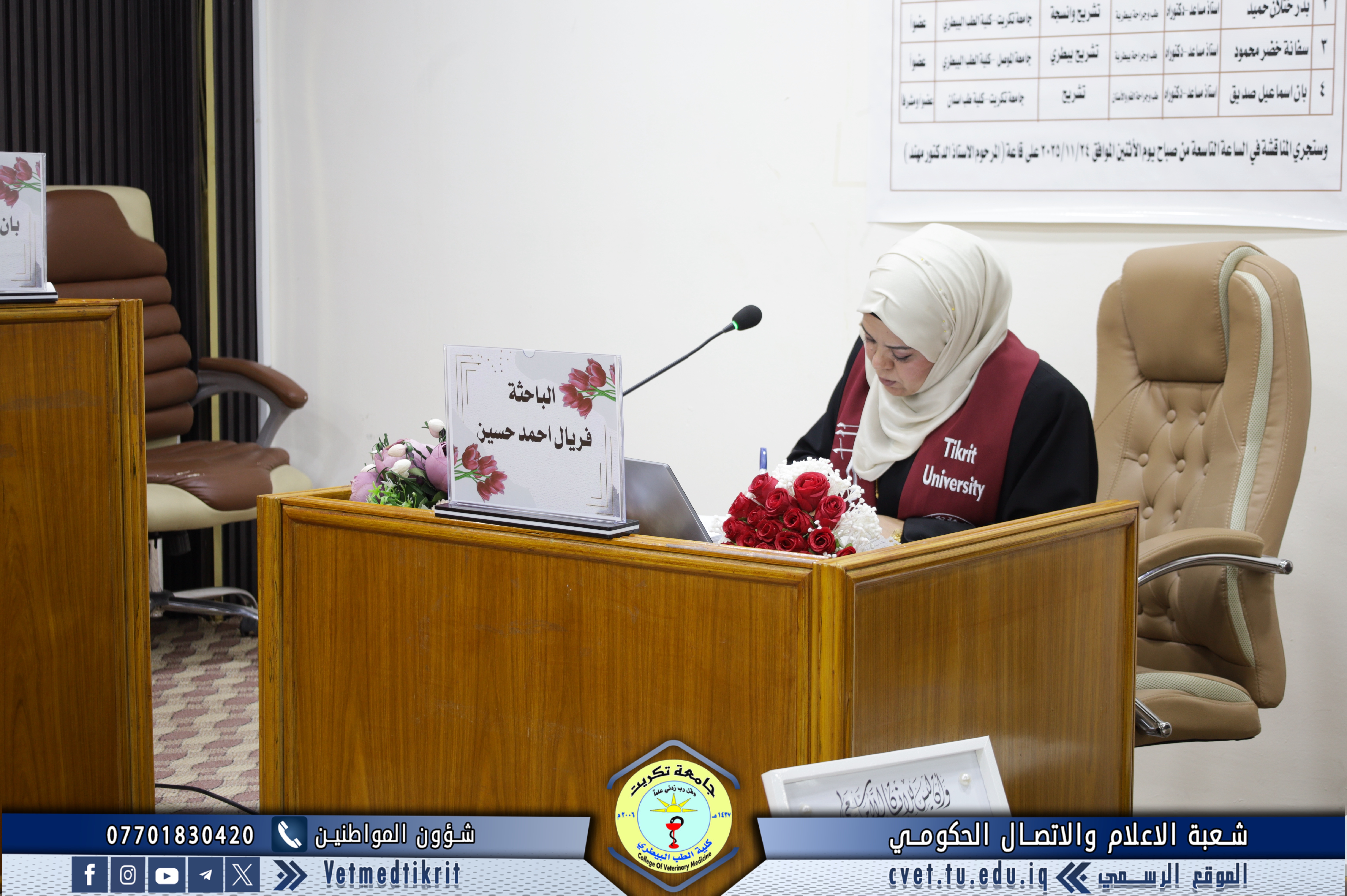
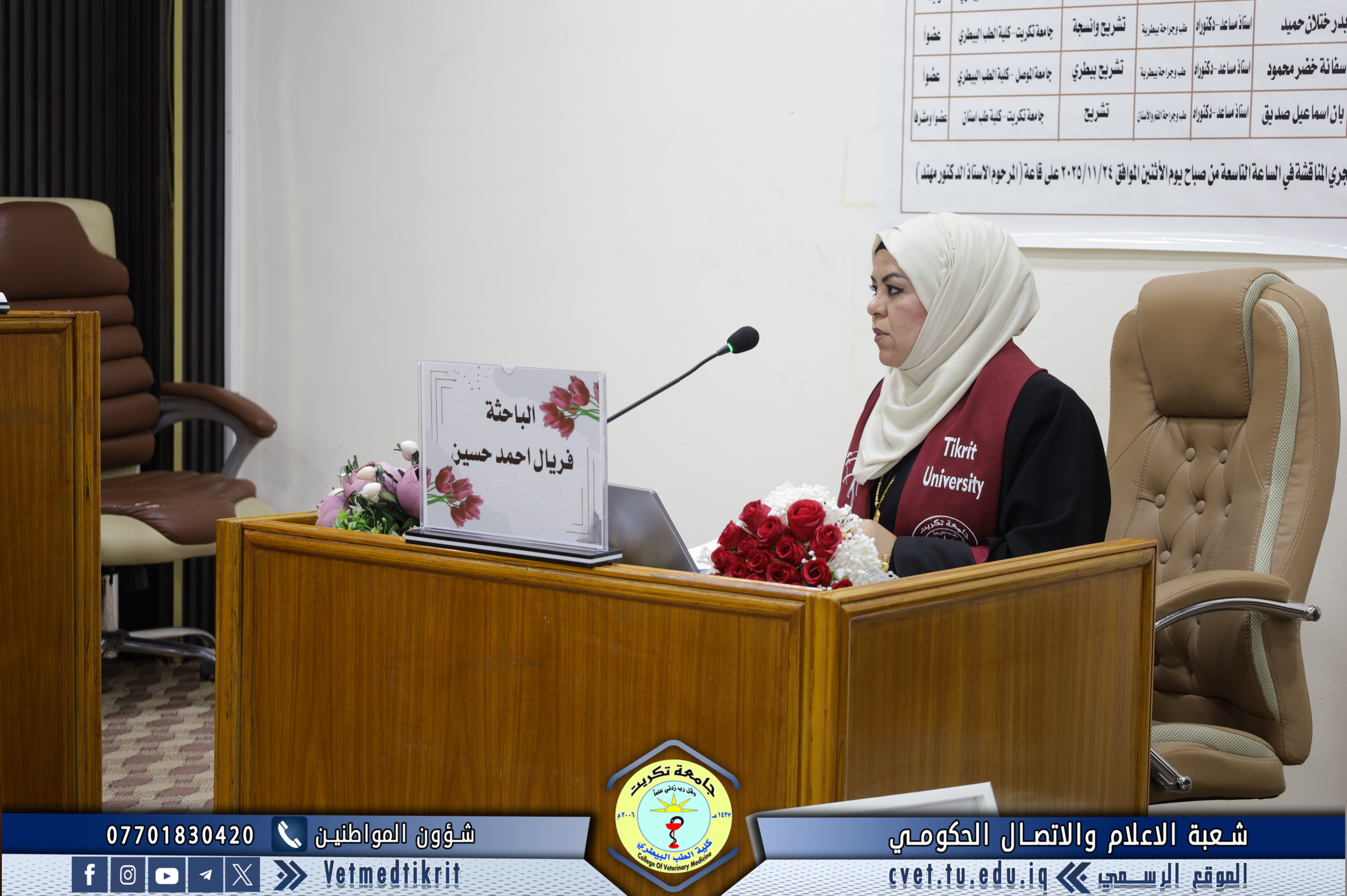
With the blessing of Almighty God, the College of Veterinary Medicine at the University of Tikrit held the defense of the master’s thesis titled
“The Effect of White Cabbage Extract on the Thyroid Gland in Adult Male Rats (A Histological Study)”
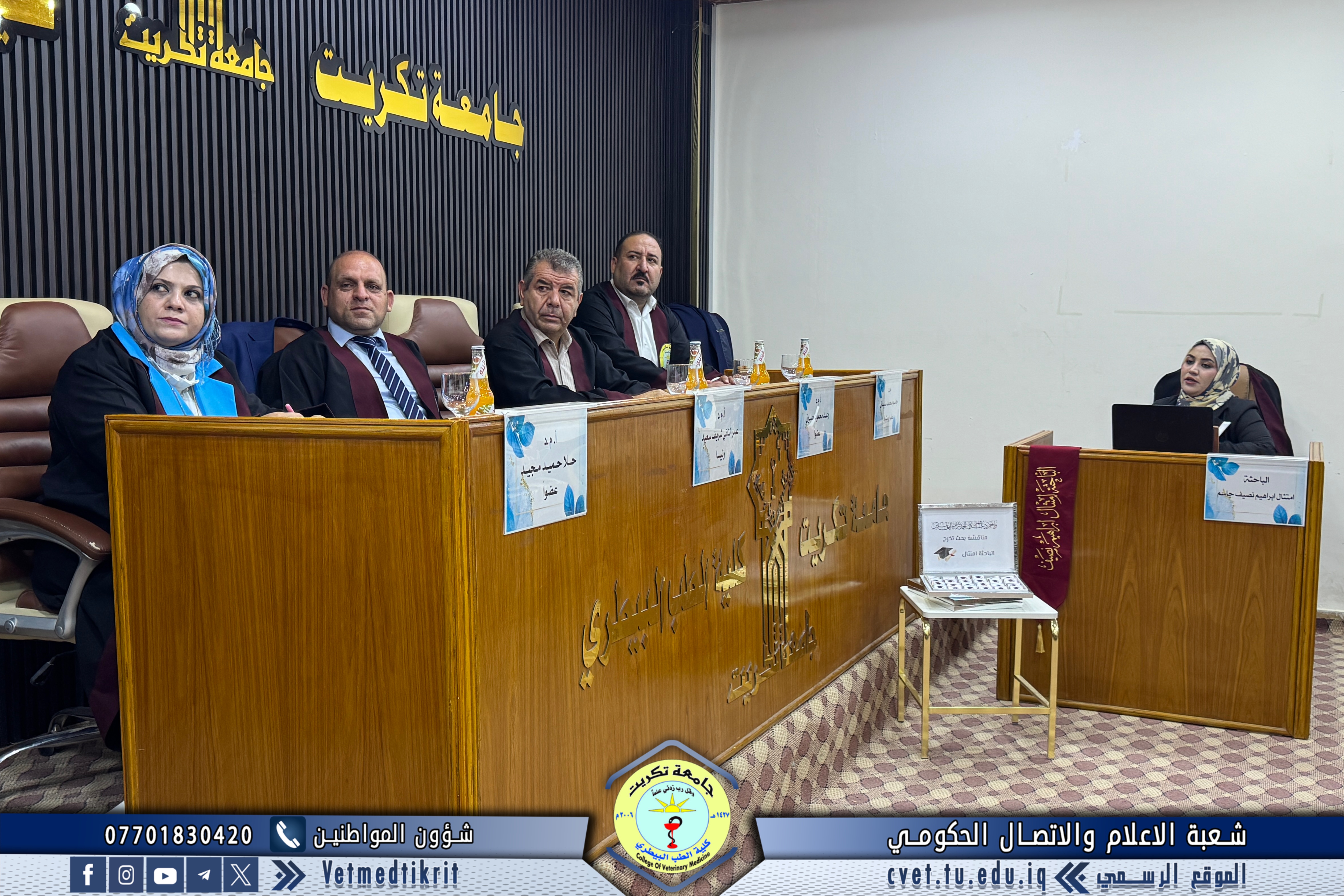
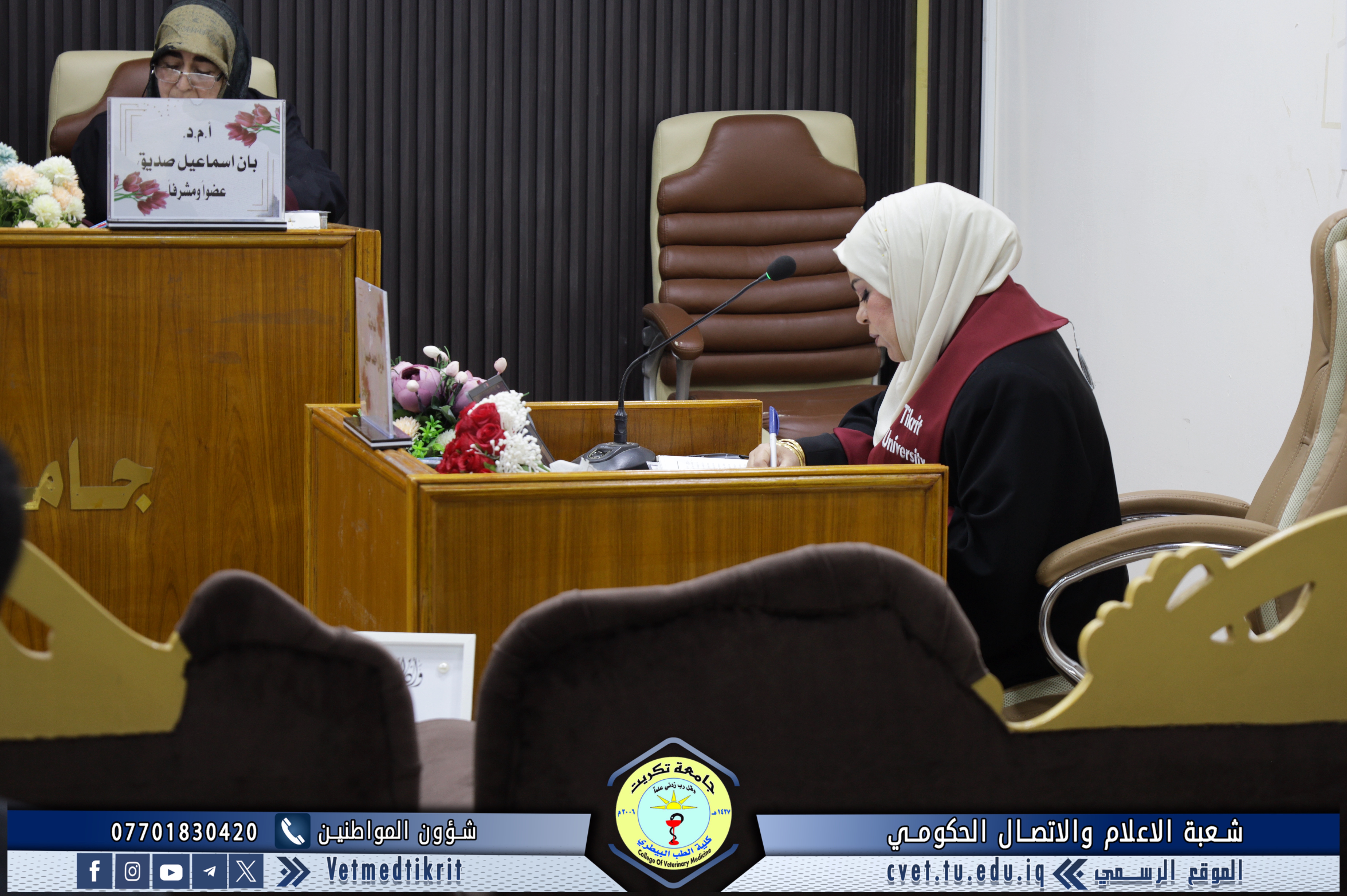
submitted by the student Feryal Ahmed Hussein, specializing in Veterinary Histology.
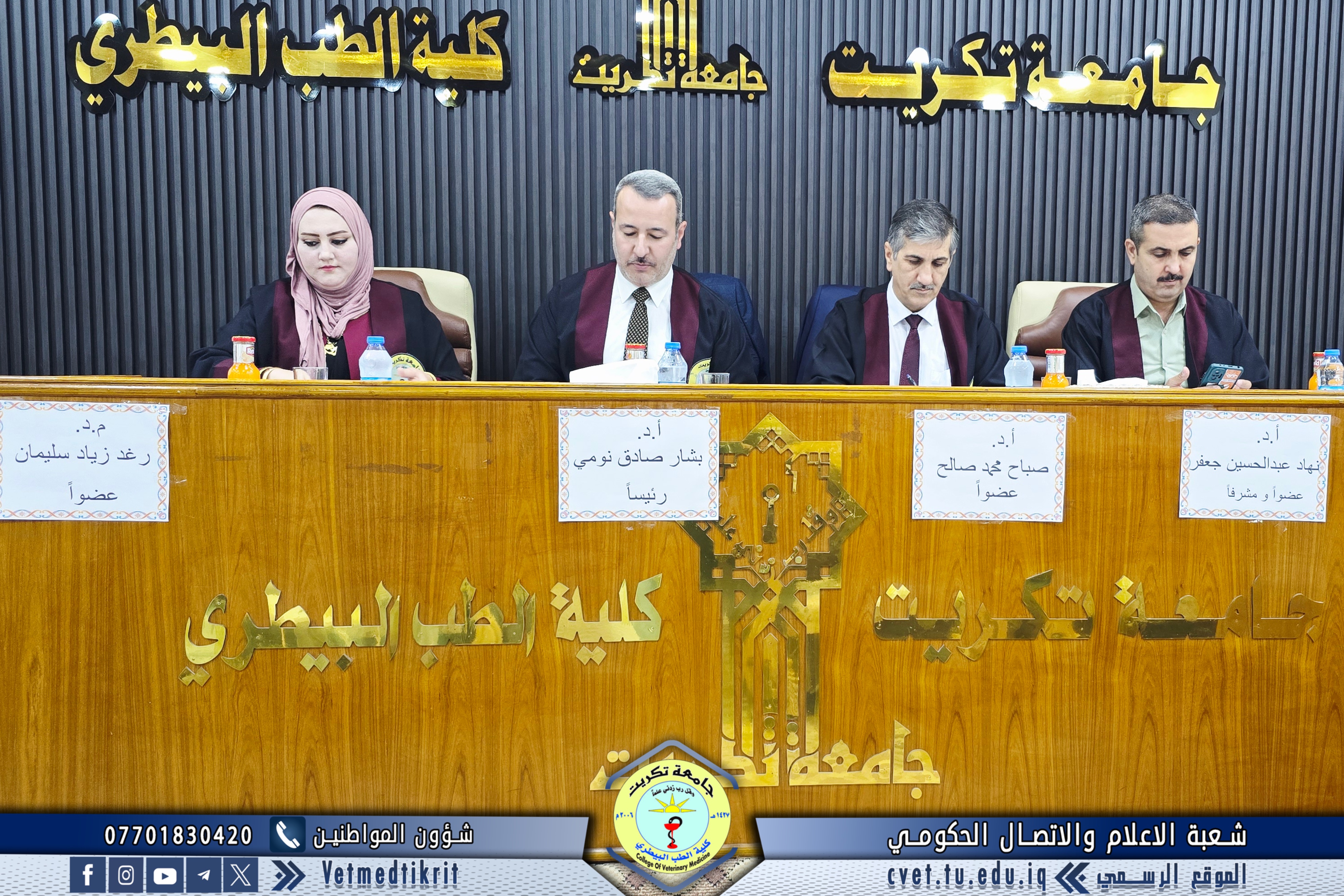
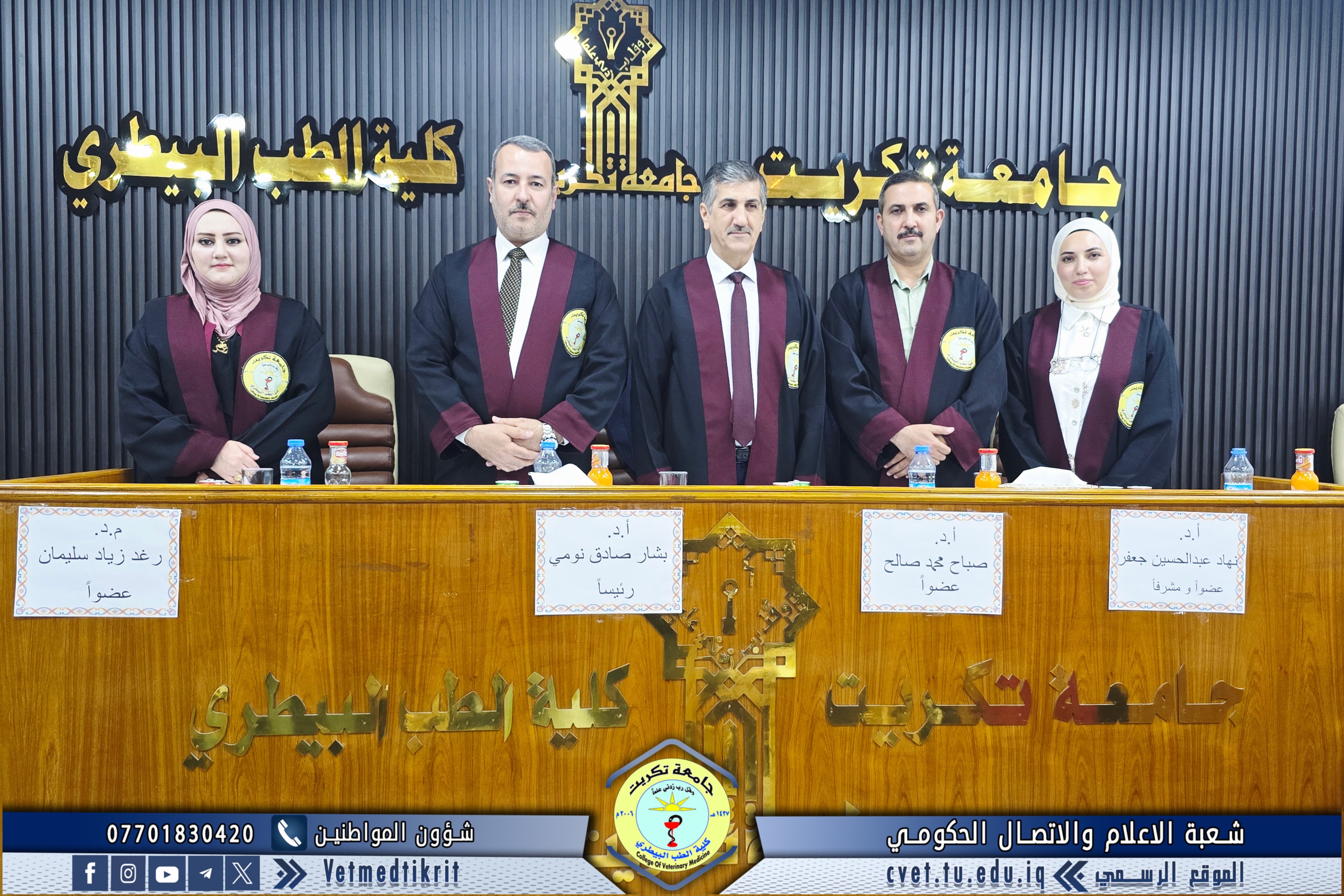
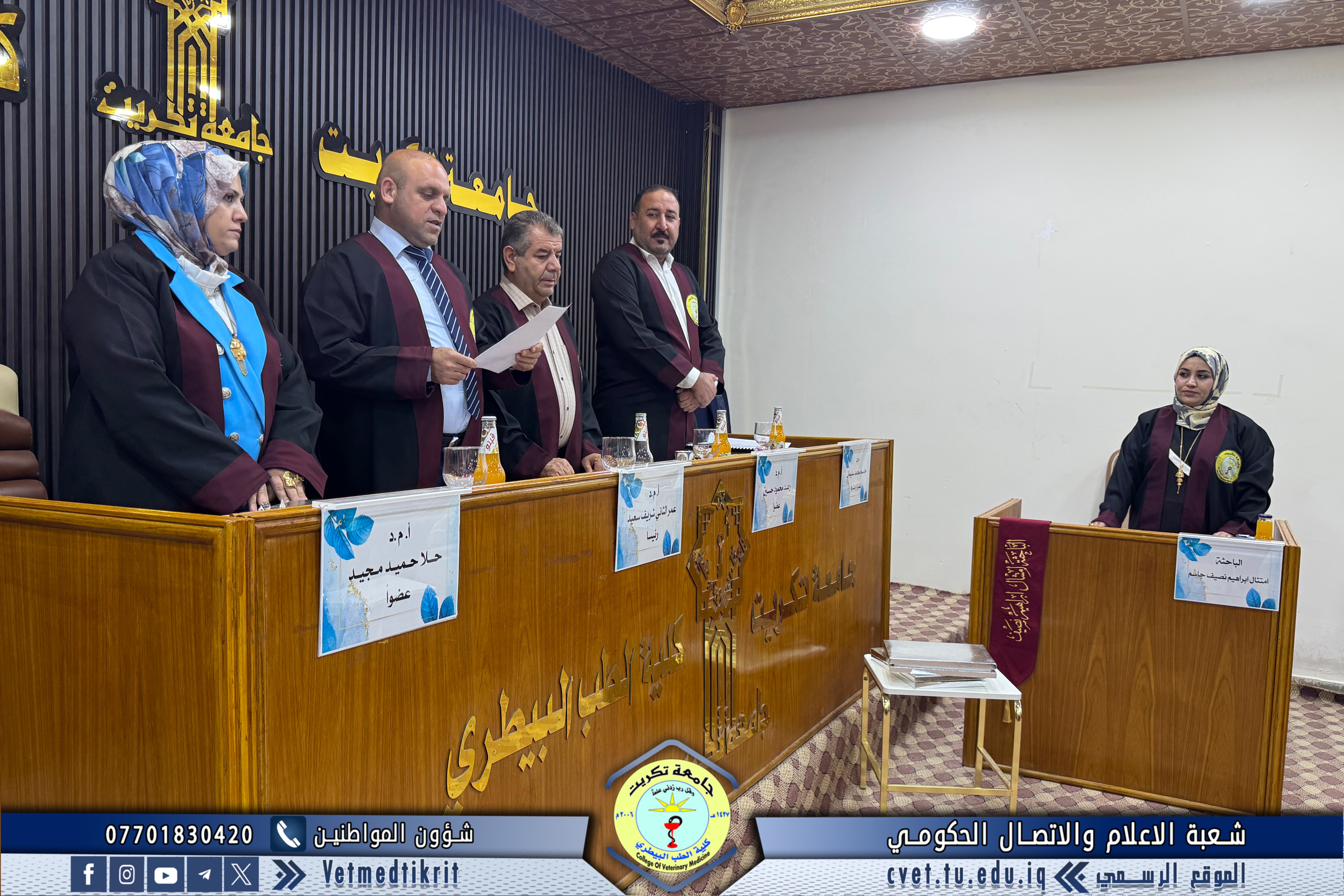
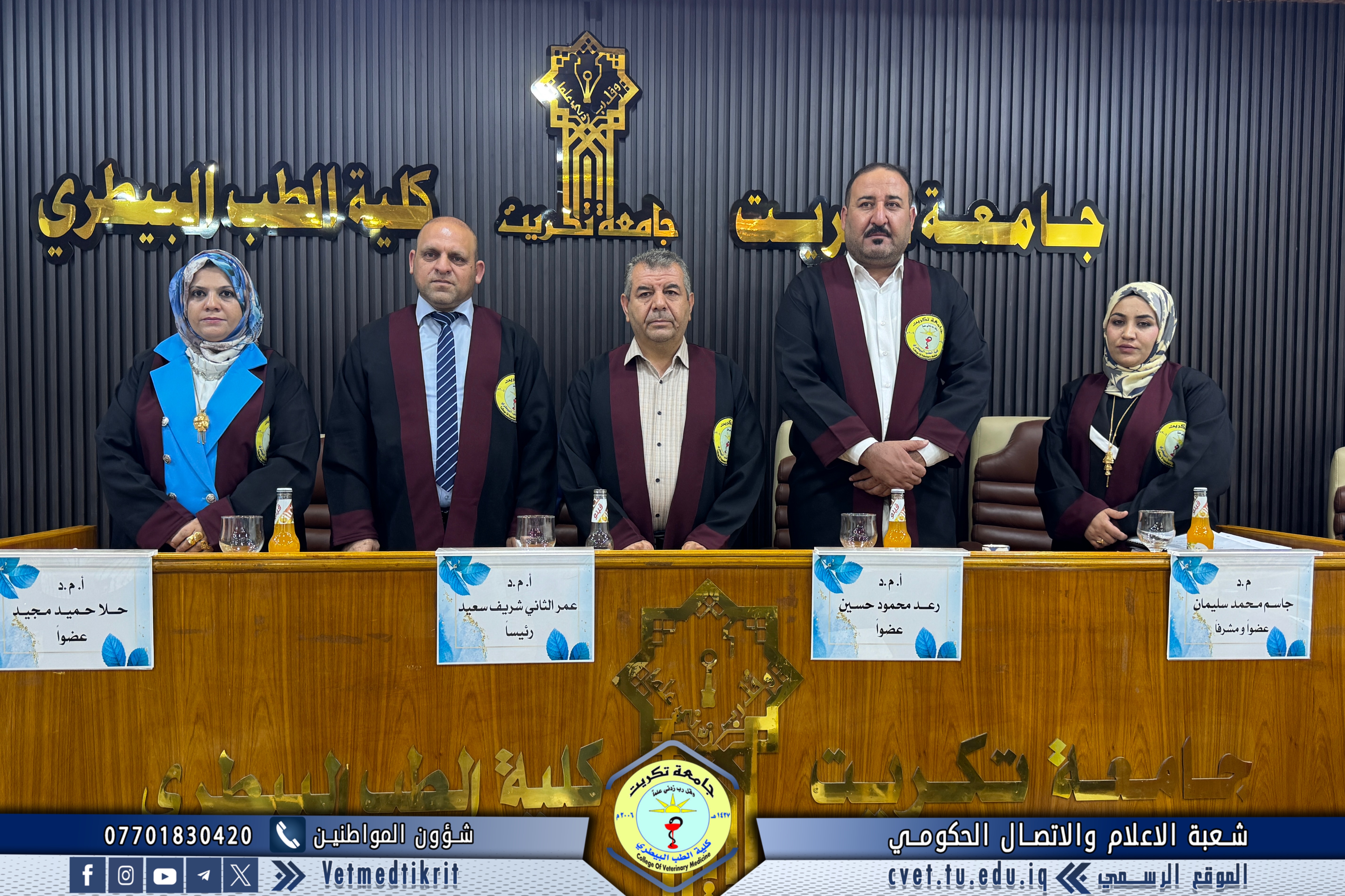
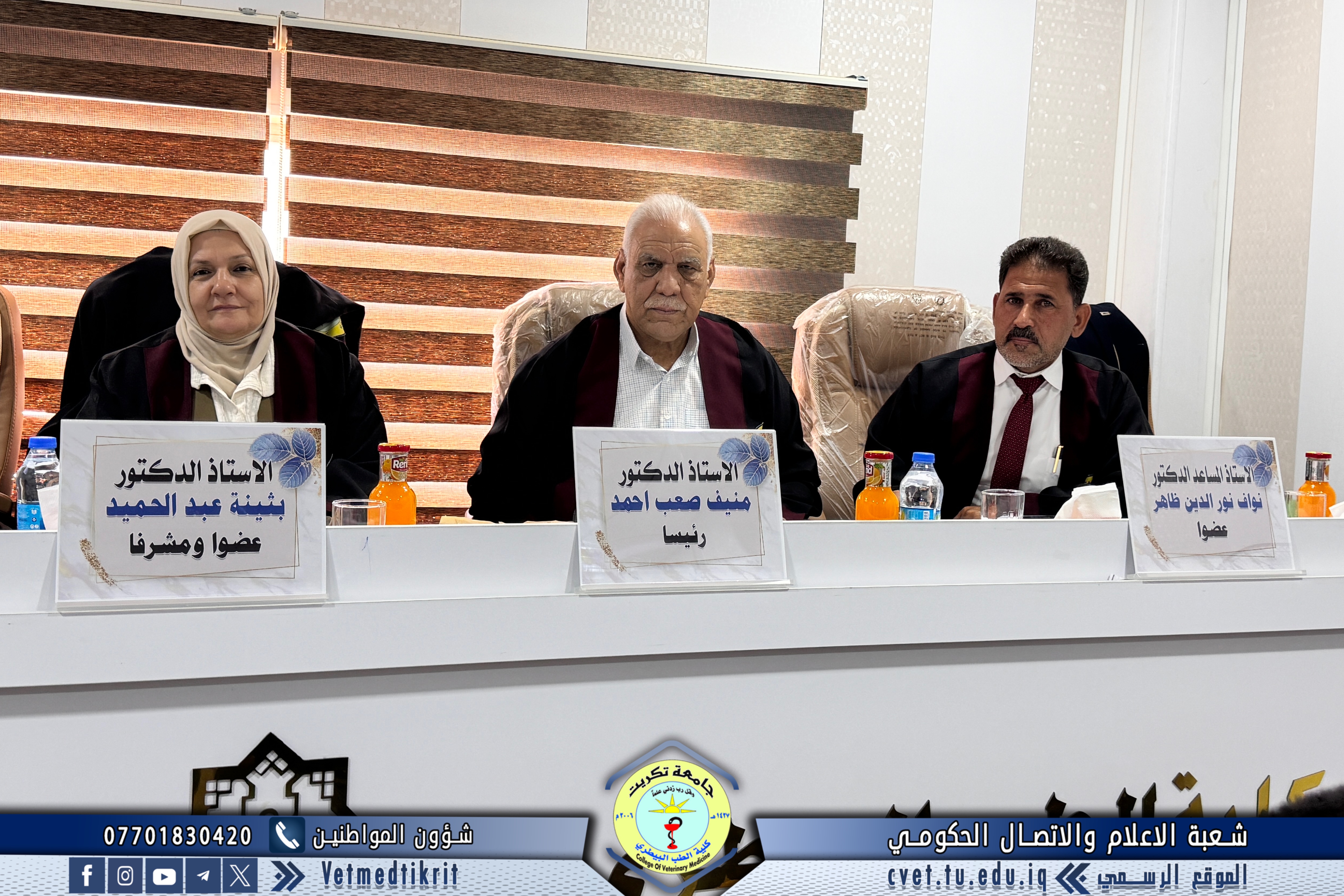
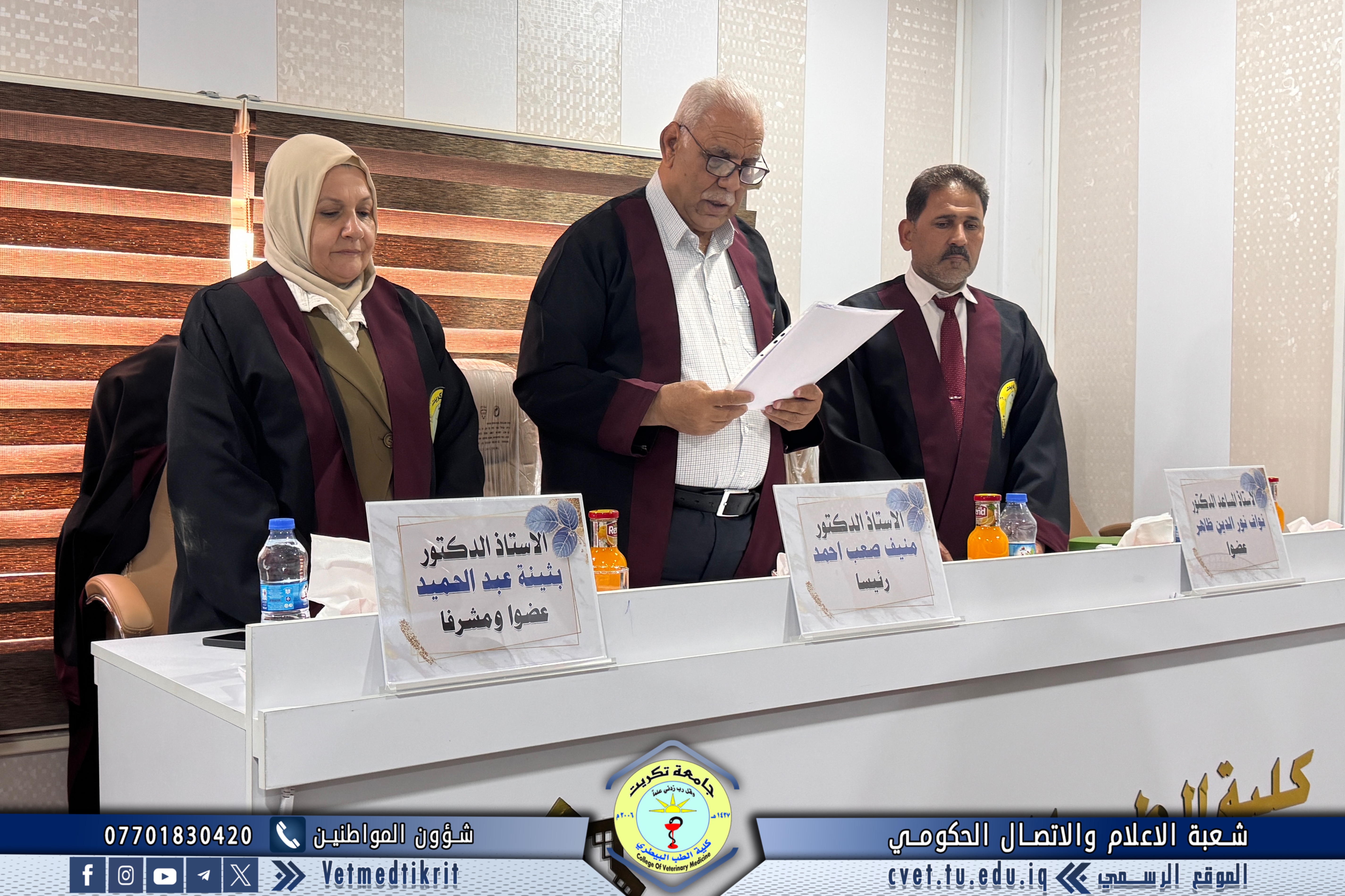
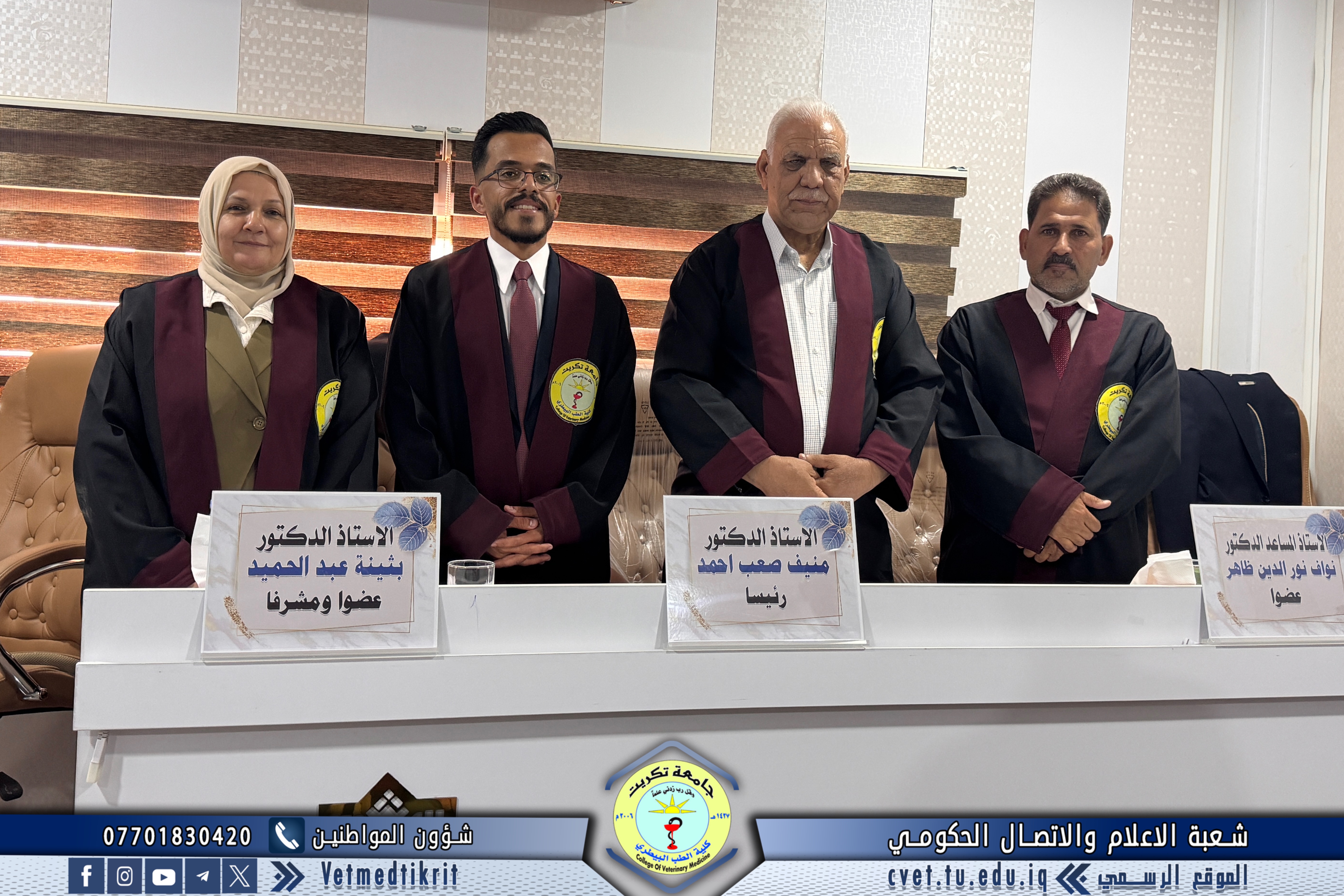
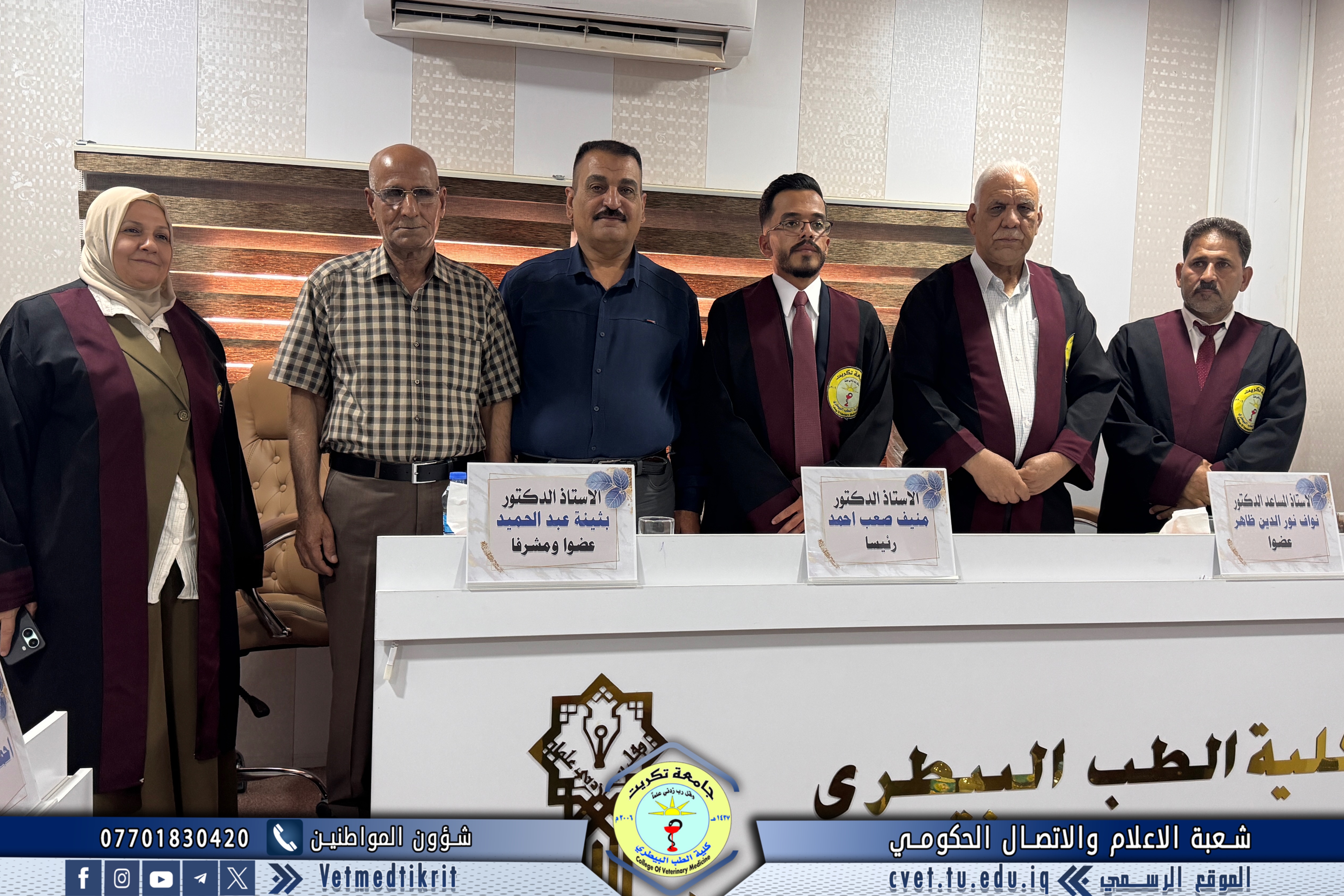
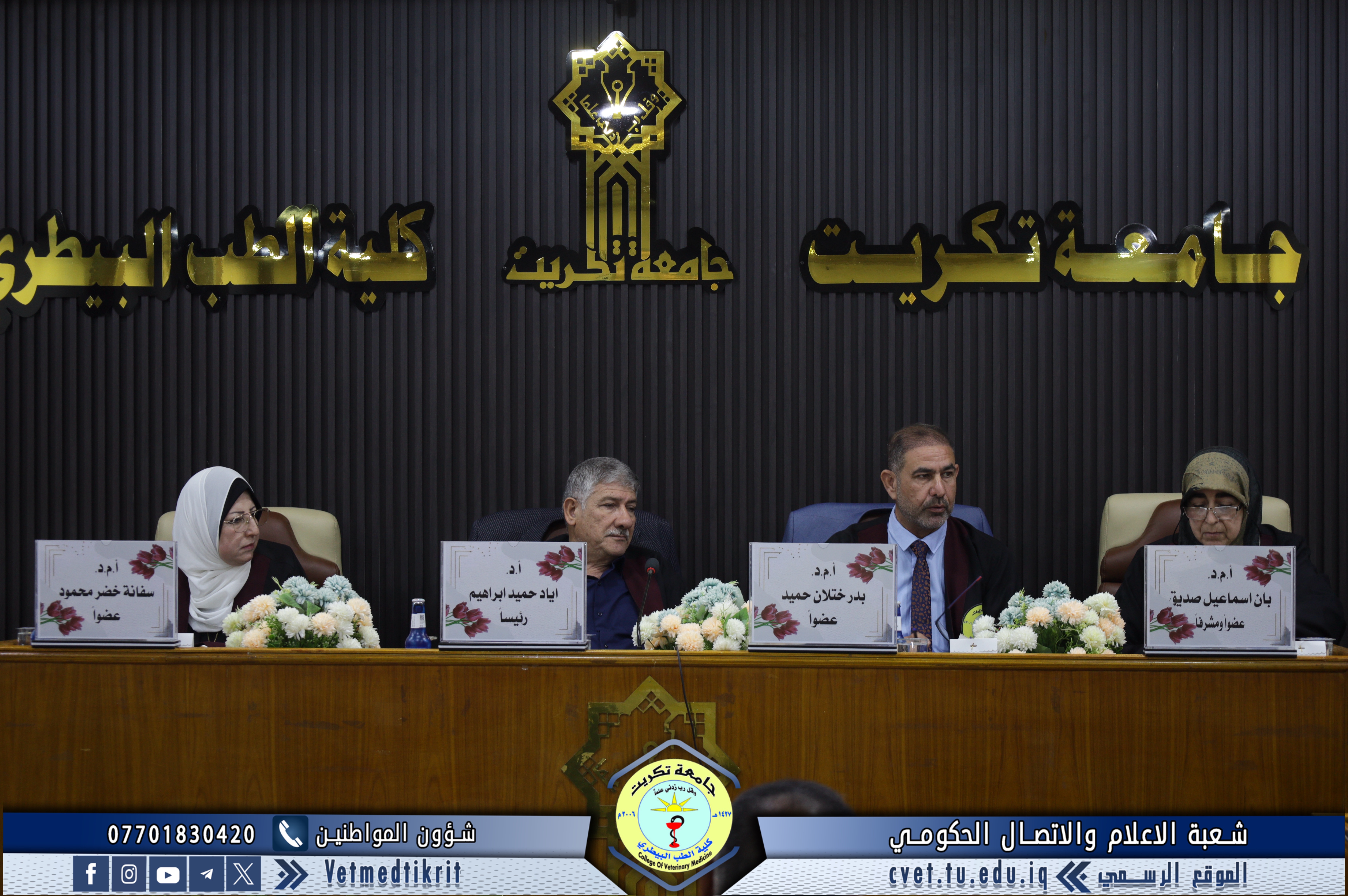
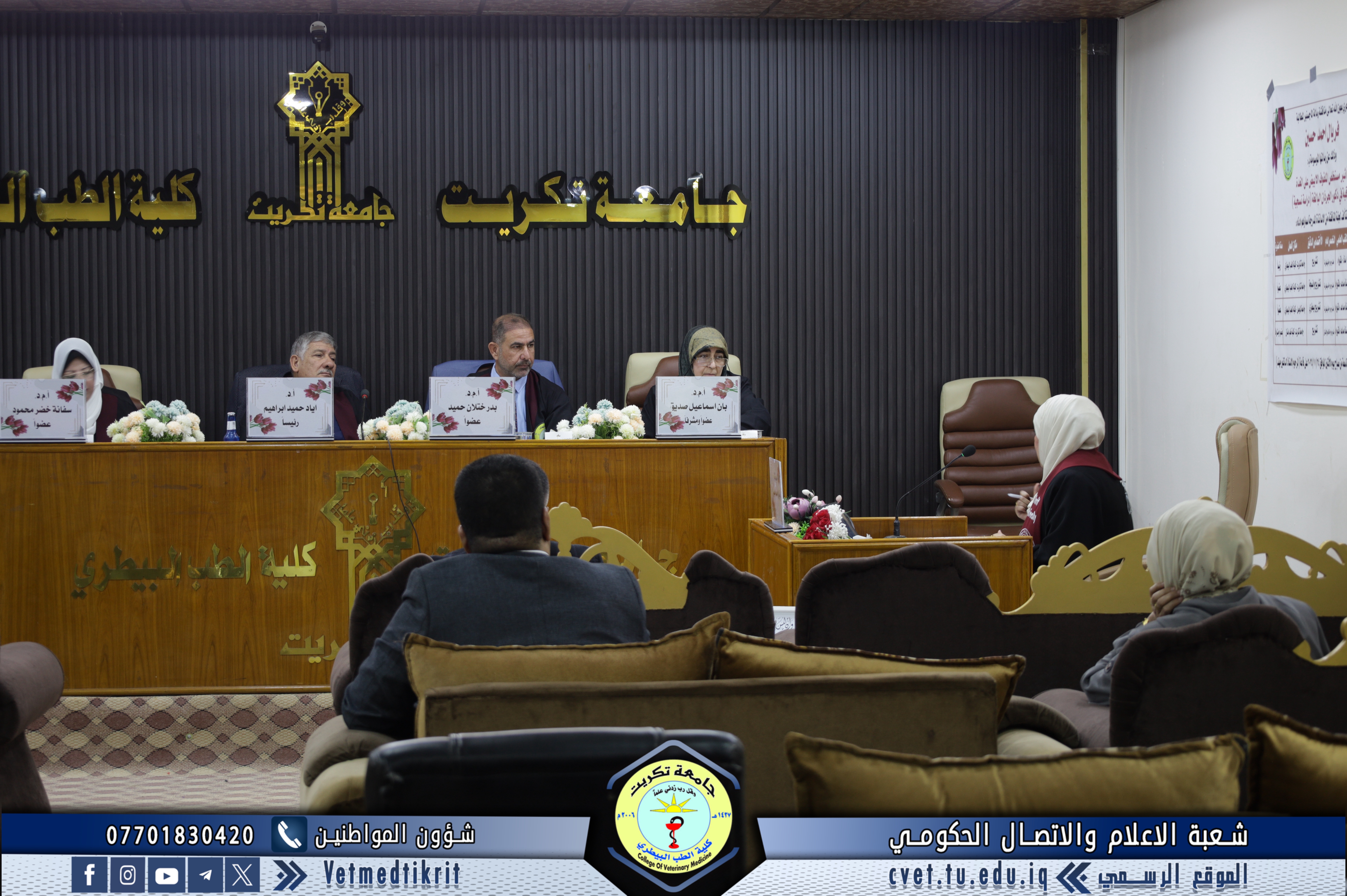
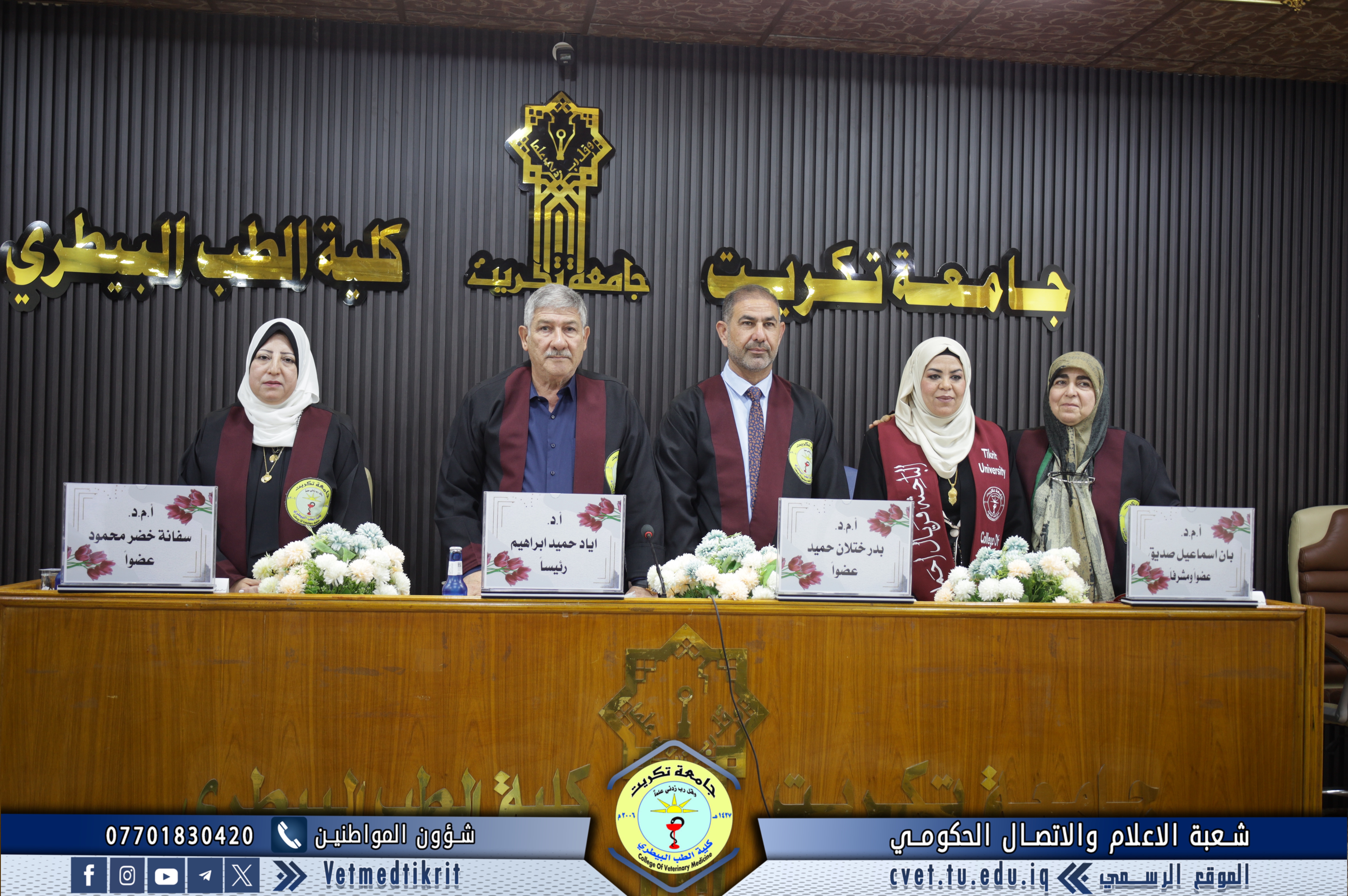
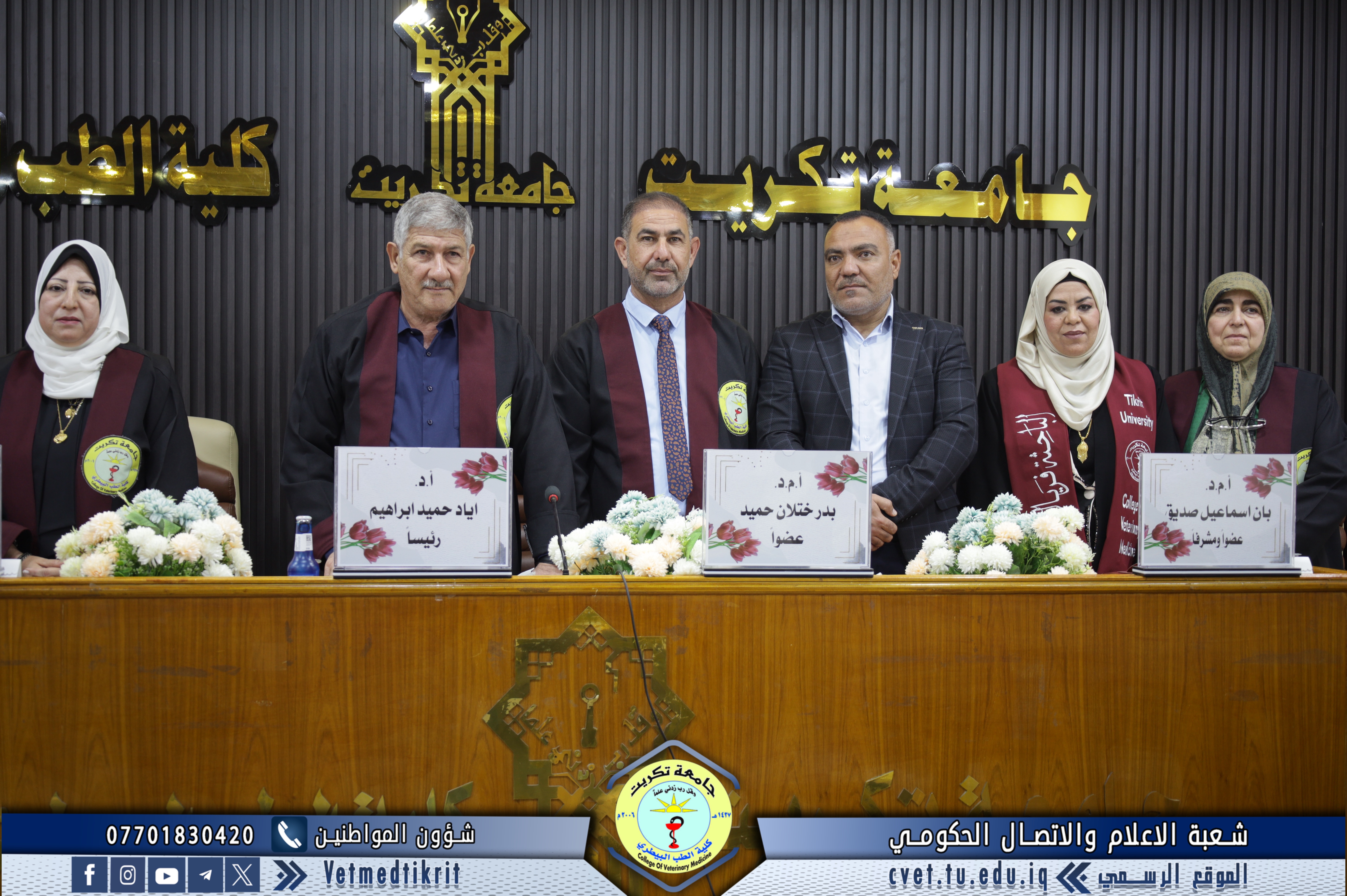
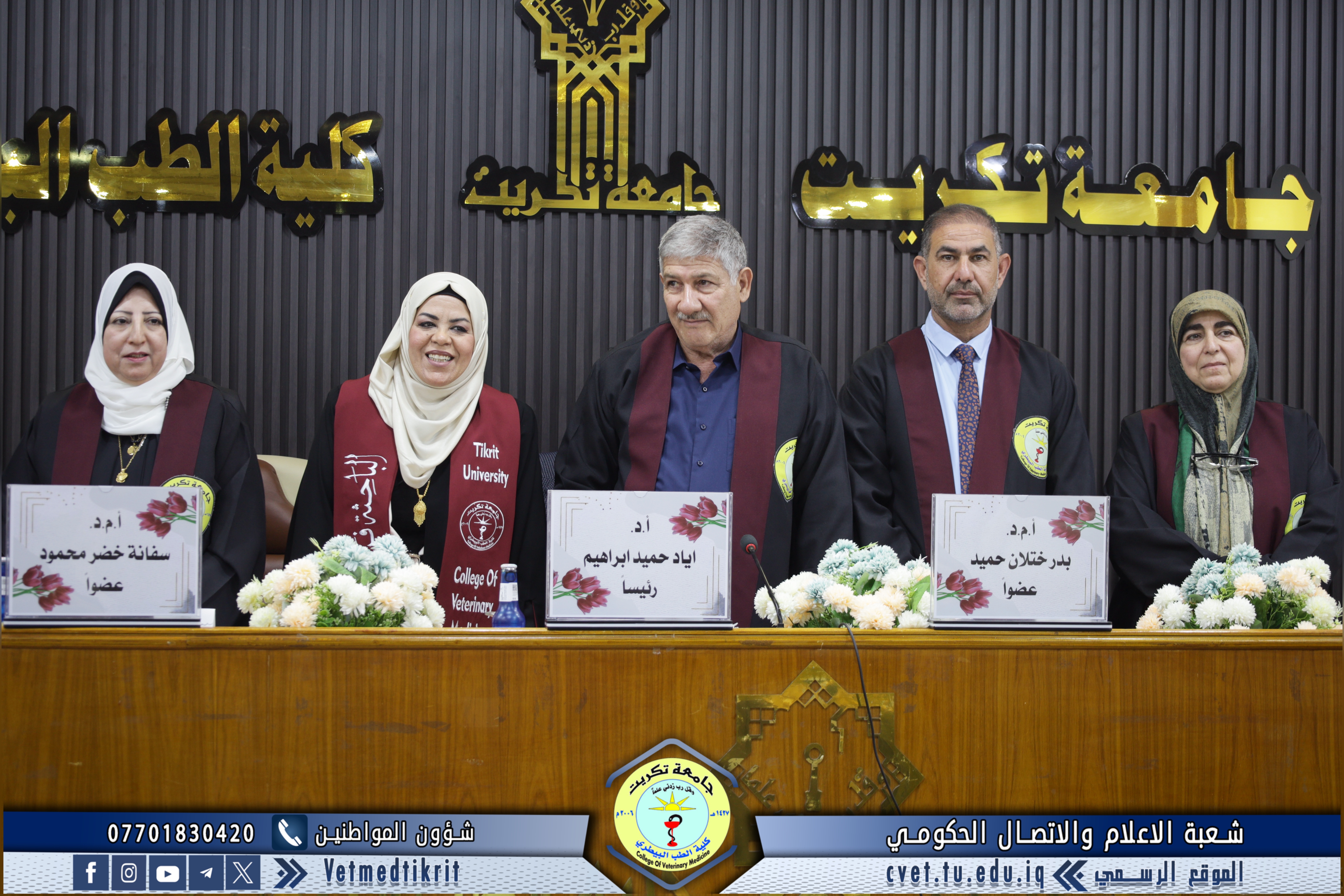
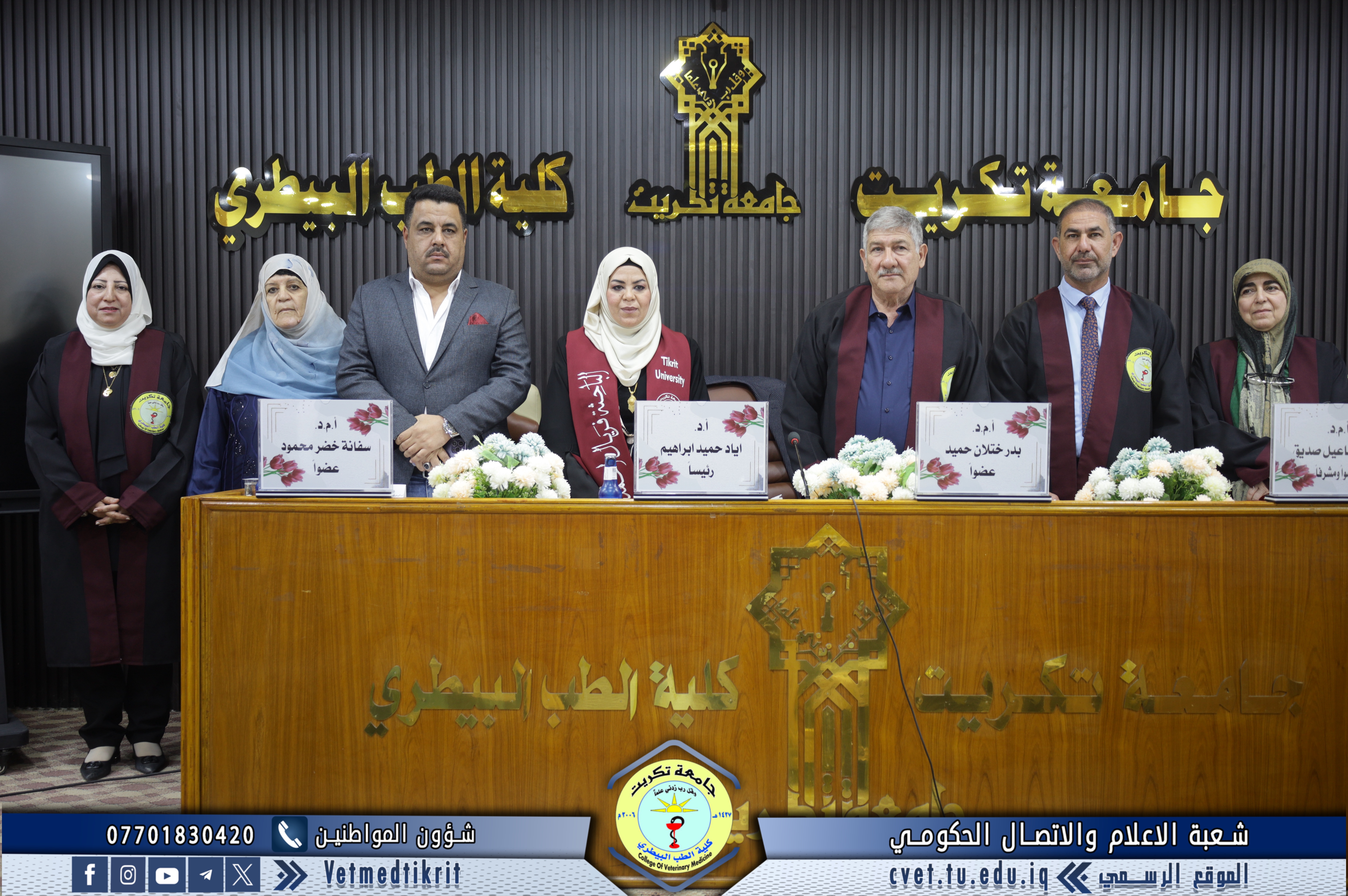
The examination committee consisted of:
-
Prof. Dr. Iyad Hameed Ibrahim / Anatomy / University of Tikrit – College of Veterinary Medicine / Chair
-
Asst. Prof. Dr. Badr Khatlan Hameed / Anatomy and Histology / University of Tikrit – College of Veterinary Medicine / Member
-
Asst. Prof. Dr. Sufana Khudhur Mahmood / Veterinary Anatomy / University of Mosul – College of Veterinary Medicine / Member
-
Asst. Prof. Dr. Ban Ismail Sidiq / Anatomy / University of Tikrit – College of Dentistry / Member and Supervisor
Thesis Abstract:
This study aims to evaluate the effect of the ethanolic extract of white cabbage on the histological structure of the thyroid gland by analyzing its histological and functional status in adult male white rats. The study was conducted in the animal house of the College of Veterinary Medicine at the University of Tikrit from September 16, 2024 to January 16, 2025.
Twenty-eight male rats weighing 250–300 g were randomly divided into three main groups.
The control group (C) consisted of four rats that received no treatment.
The experimental group (A) consisted of twelve rats divided into three subgroups (A1, A2, A3), each containing four rats, which received 100 mg/kg of the extract three times per week for one, two, and four months respectively.
Group (B) also consisted of twelve rats divided into subgroups (B1, B2, B3), each containing four rats, and received the same dose daily for one, two, and four months respectively.
At the end of the treatment period, serum levels of T3, T4, and TSH were measured using the ELISA technique. Thyroid tissues were also subjected to histological examination using Hematoxylin and Eosin, Periodic Acid–Schiff (PAS), and Masson’s Trichrome stains. Morphological changes were evaluated by measuring follicular diameter, epithelial cell height, and the number of cells per follicle.
Histological results in the control group showed a normal structure, with follicles filled with homogeneous colloid, cuboidal epithelium, and intact follicular cells.
In contrast, treated groups exhibited progressive pathological alterations. Subgroups A1 and B1 showed follicles containing heterogeneous colloid, vacuolar degeneration of follicular cells, desquamation in some follicles, and moderate epithelial flattening.
With longer treatment durations in A2 and B2, the severity increased to include hemorrhage, follicular enlargement, and colloid depletion.
The most severe changes appeared in A3 and B3, which demonstrated irregular follicles, cellular atrophy, necrosis, and pronounced fibrosis. Masson’s Trichrome staining revealed collagen deposition in the interfollicular connective tissue, especially in group B3, indicating tissue fibrosis. PAS staining showed abnormal reactions with dense colloid accumulation, suggesting follicular inactivity and reduced hormone production.
Quantitative histological measurements supported these findings:
Follicular diameters in B2 (167.9 ± 7.7 µm) and A2 (155.8 ± 8.1 µm) were significantly larger than those of the control group (103.2 ± 8.6 µm), indicating colloid buildup and follicular hypertrophy.
In A3 and B3, follicular diameters decreased markedly, reflecting degenerative collapse.
Epithelial height declined significantly from 8.64 ± 0.7 µm in the control to 4.2 ± 0.12 µm in B3, indicating reduced metabolic activity and cellular atrophy.
The number of cells per medium and large follicle increased in A2, A3, and B2, suggesting hyperplastic activity.
Inflammatory responses, such as leukocytic infiltration, were observed in A2, B2, and B3, with extensive fibrosis particularly in B3.
Hormonal analysis revealed a significant (P ≤ 0.05) decrease in T3 and T4 levels in the treated groups compared to the control, especially in group B3, where T3 reached 1.22 ± 0.03 ng/mL and T4 reached 5.64 ± 0.19 µg/dL. This was accompanied by a compensatory elevation in TSH, which reached its highest value (23.15 ± 0.12 µIU/mL) in group B3. These hormonal disturbances confirmed hypothyroidism induced by the extract.
The study concludes that white cabbage has the potential to cause thyroid gland damage and that long-term consumption may adversely affect thyroid function.
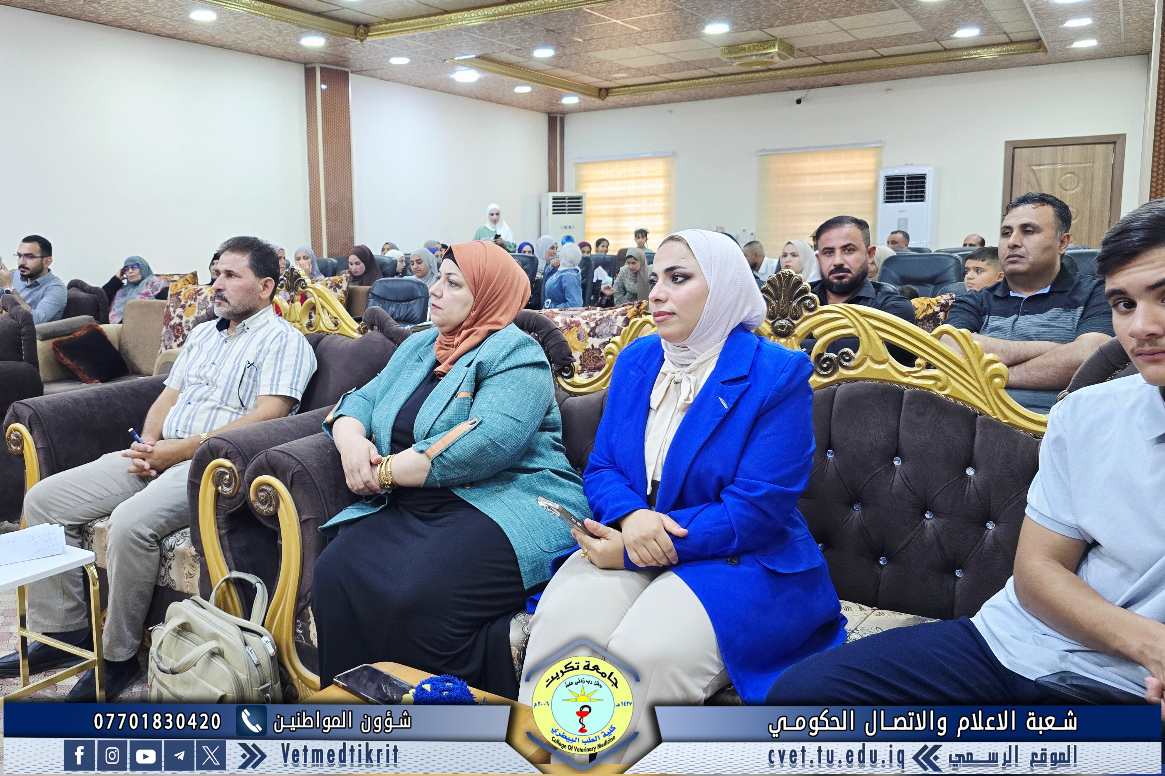
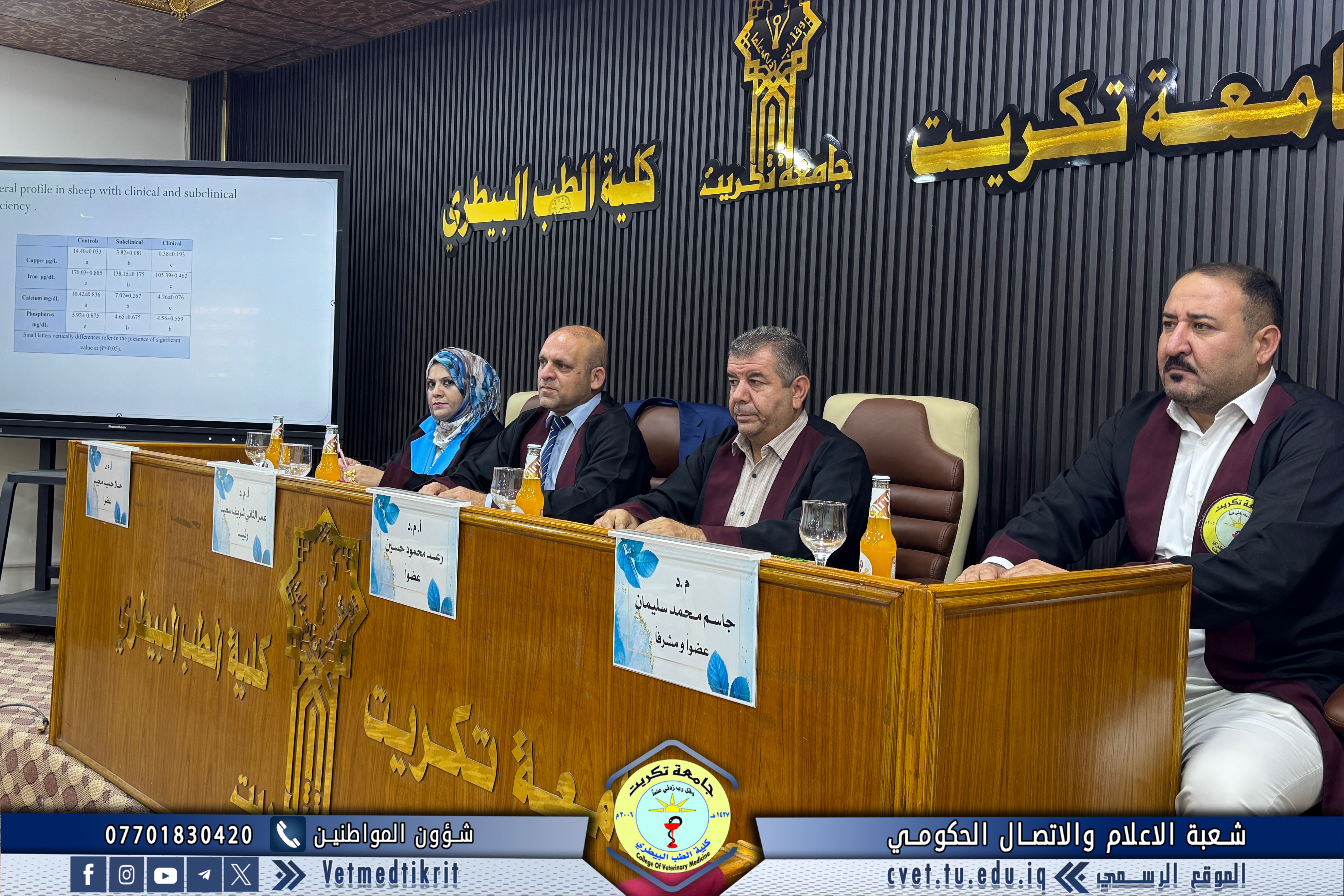
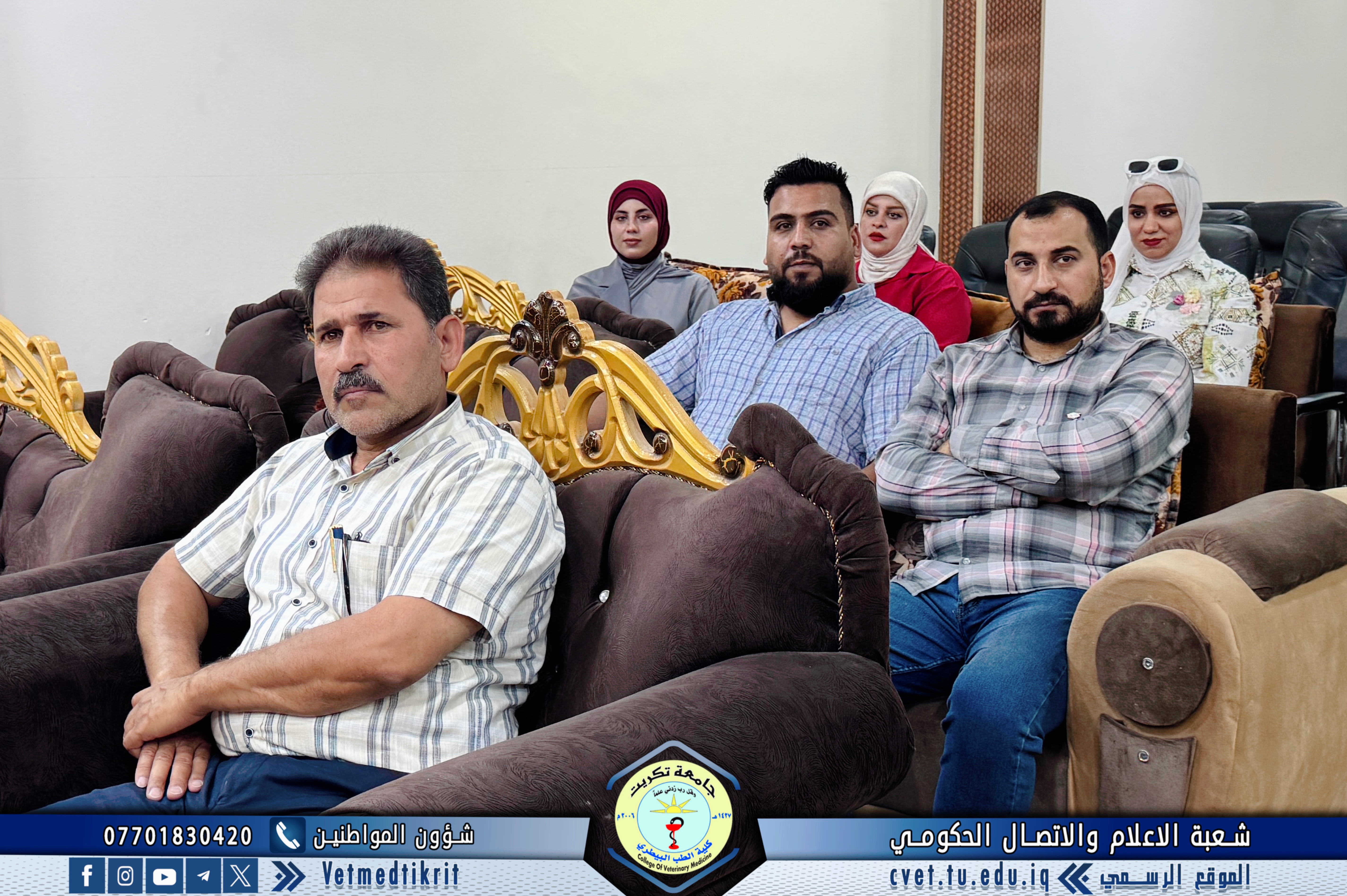
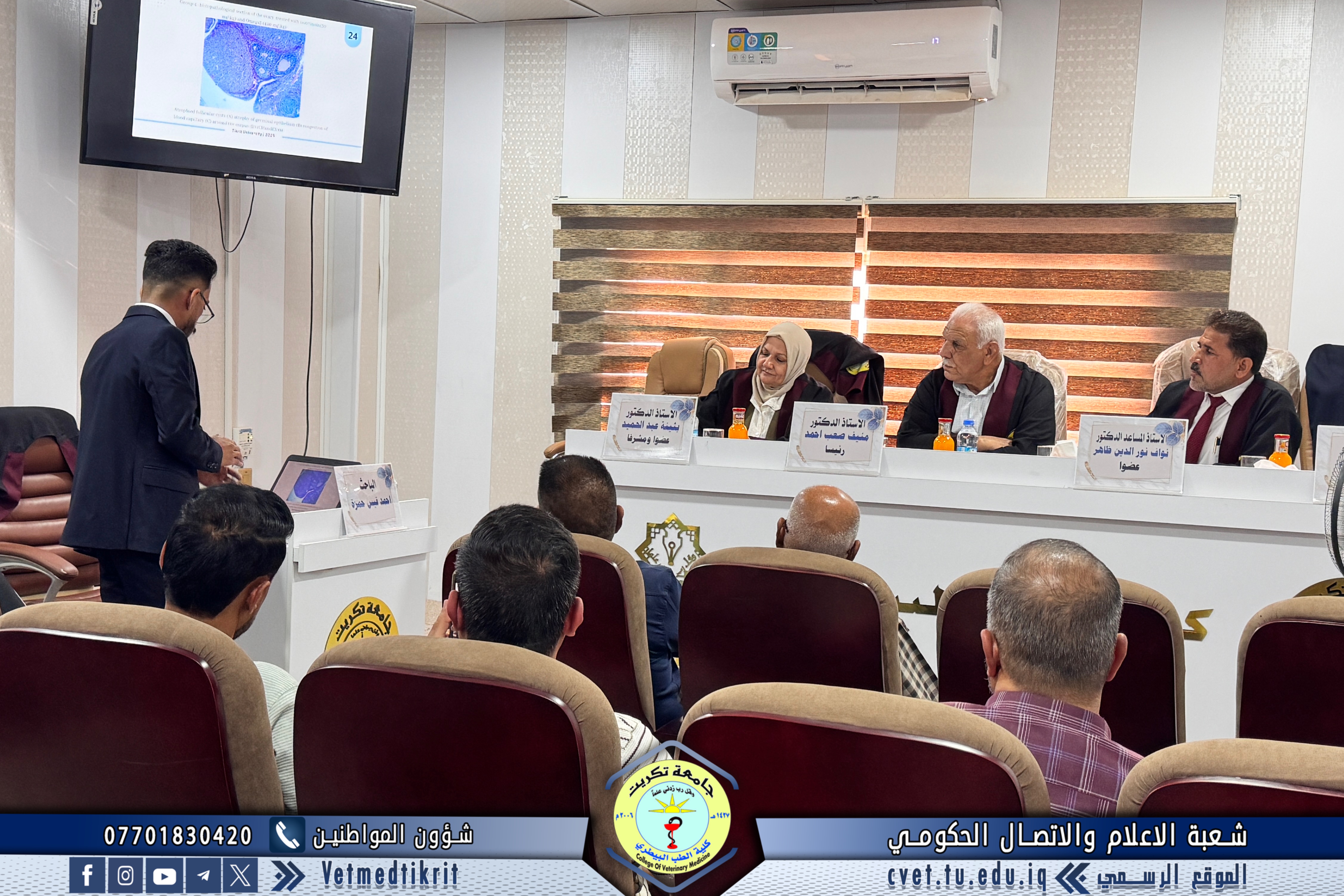
The defense session, held in Dr. Muhannad Maher Hall at the College of Veterinary Medicine, was attended by several faculty members and a number of students