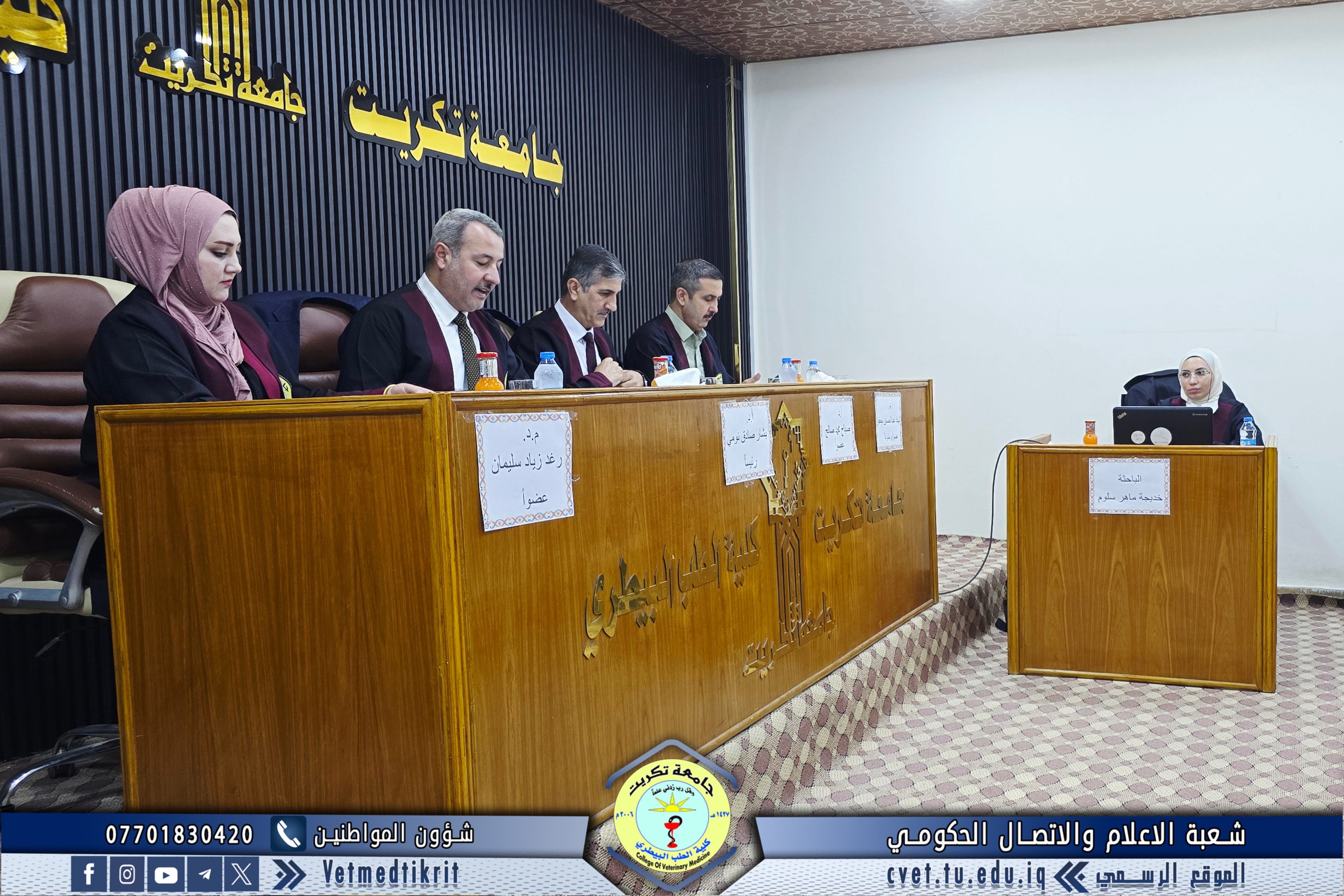
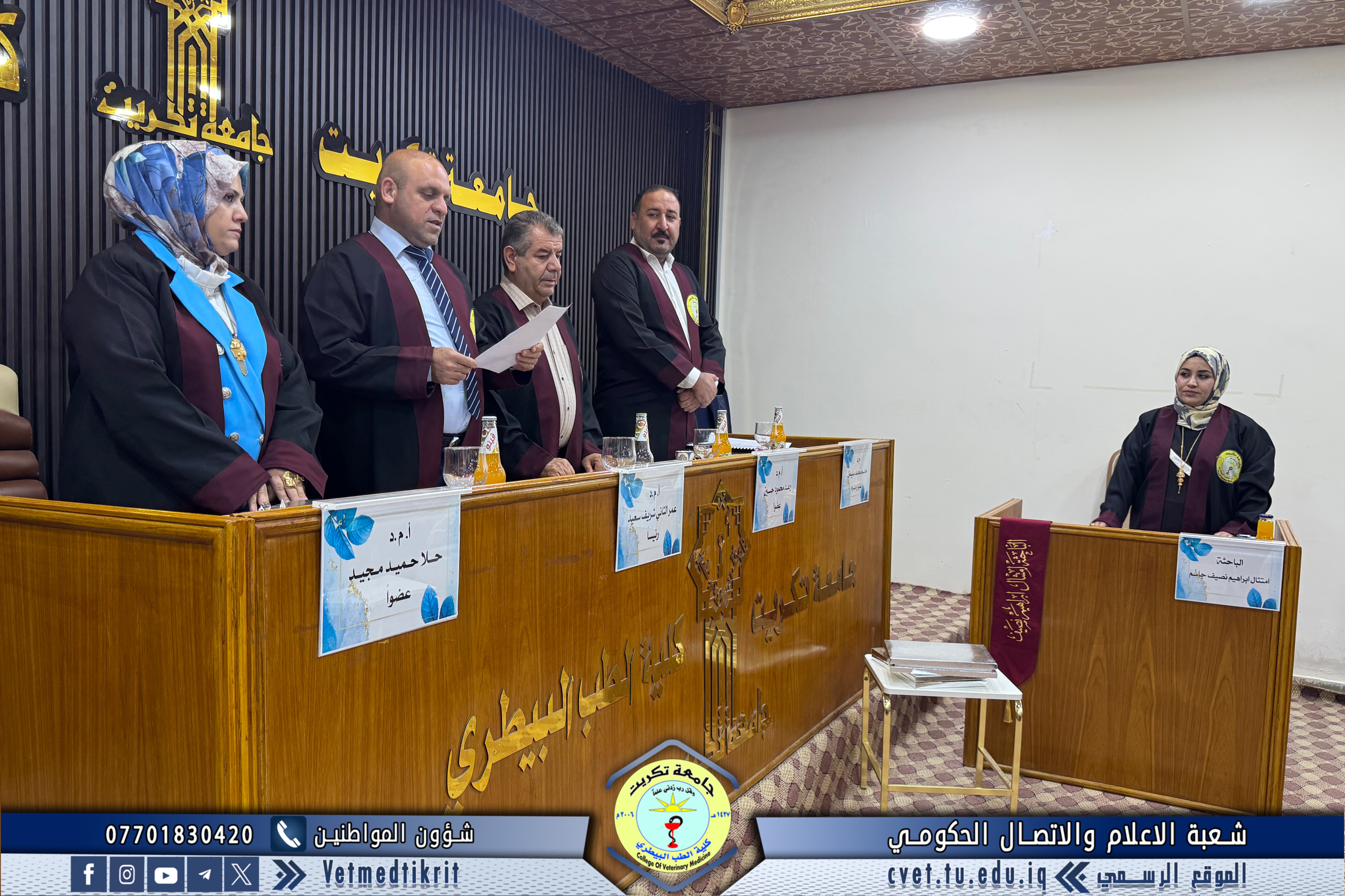
With the grace of God, the College of Veterinary Medicine at the University of Tikrit conducted the defense of the master’s thesis entitled “Anatomical and Histological Structure of the Liver and the Biliary Duct System in the Seagull and Local Pigeon: A Comparative Study” submitted by the student Du’aa Abdullah Khudhr, specializing in Veterinary Histology.
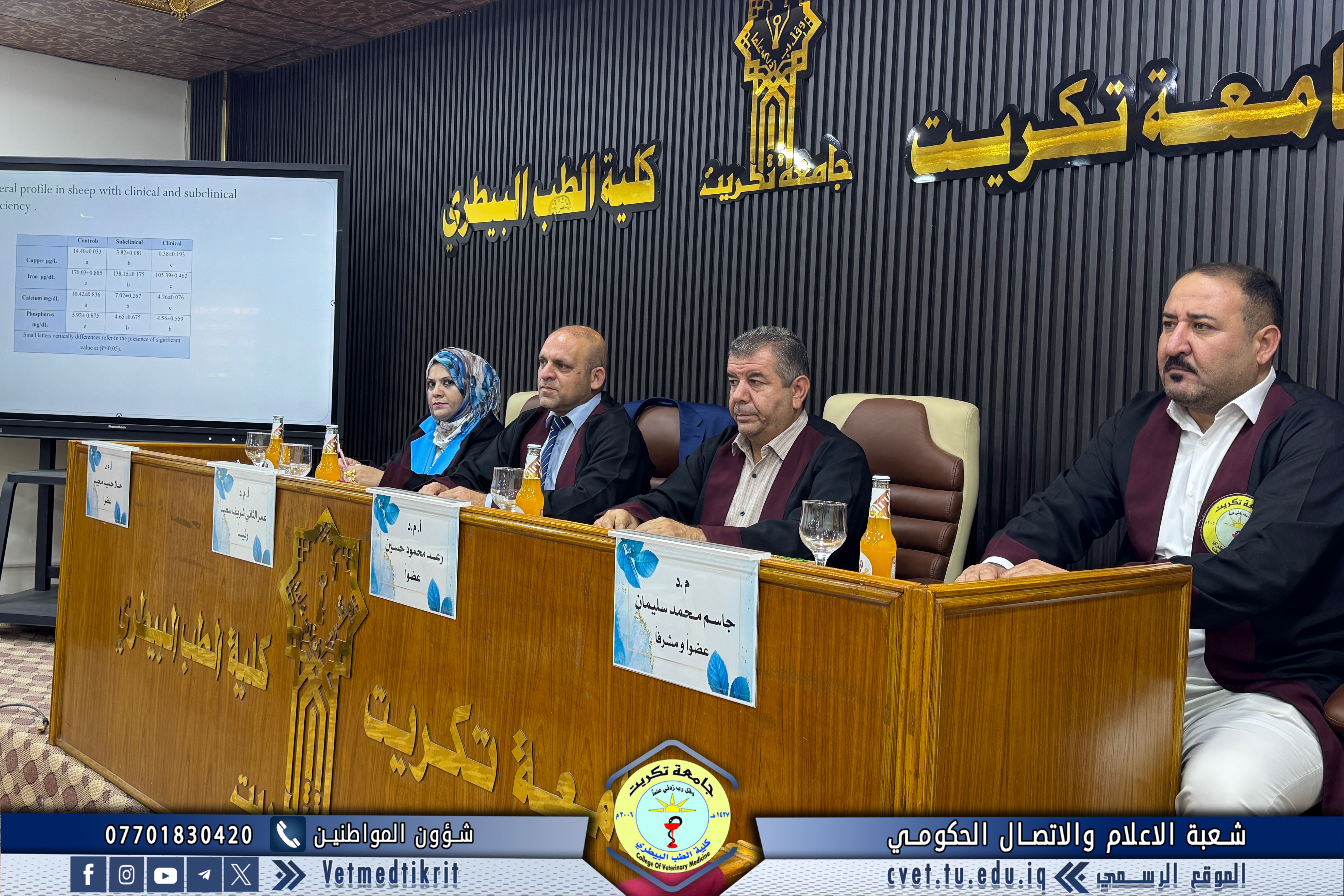
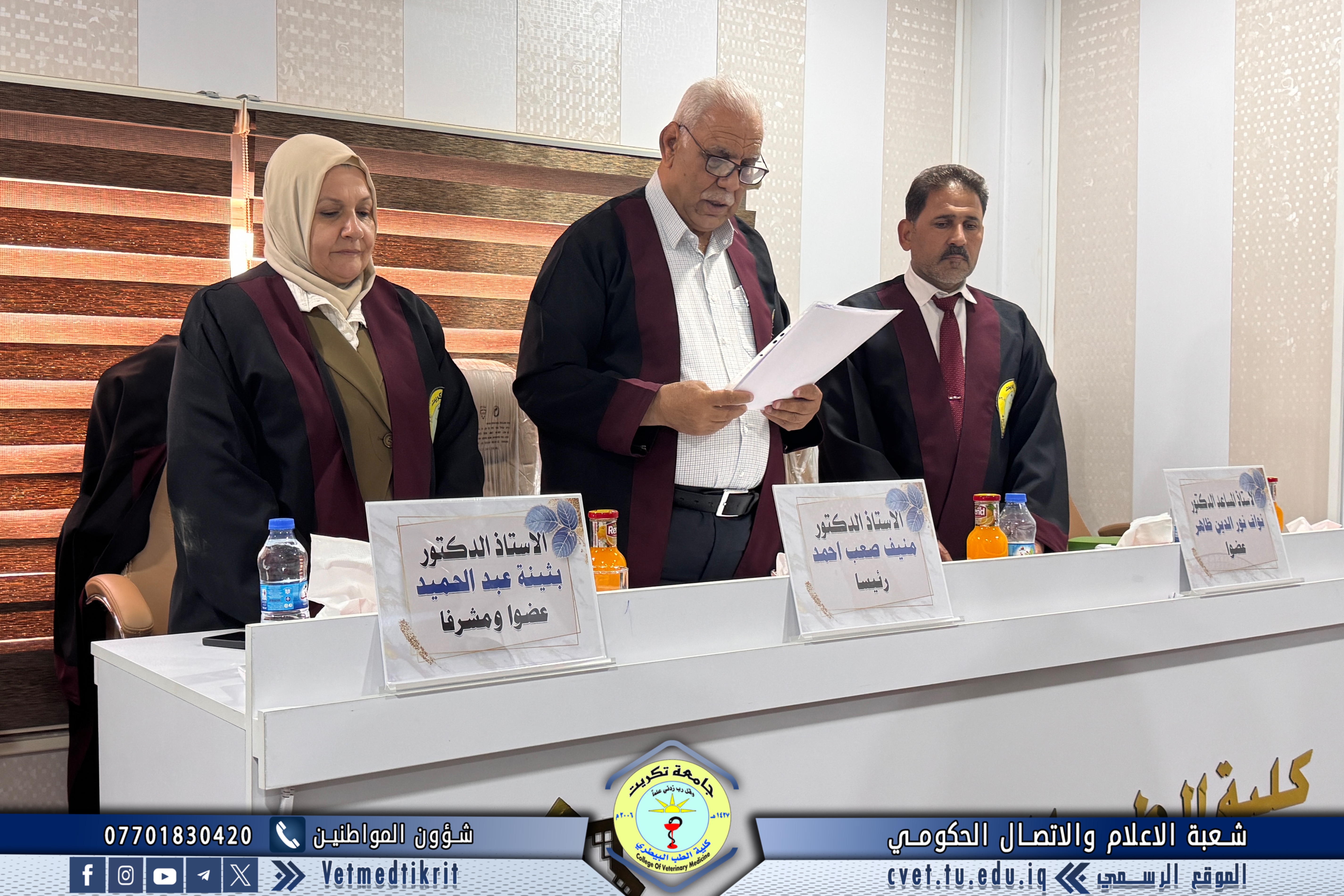
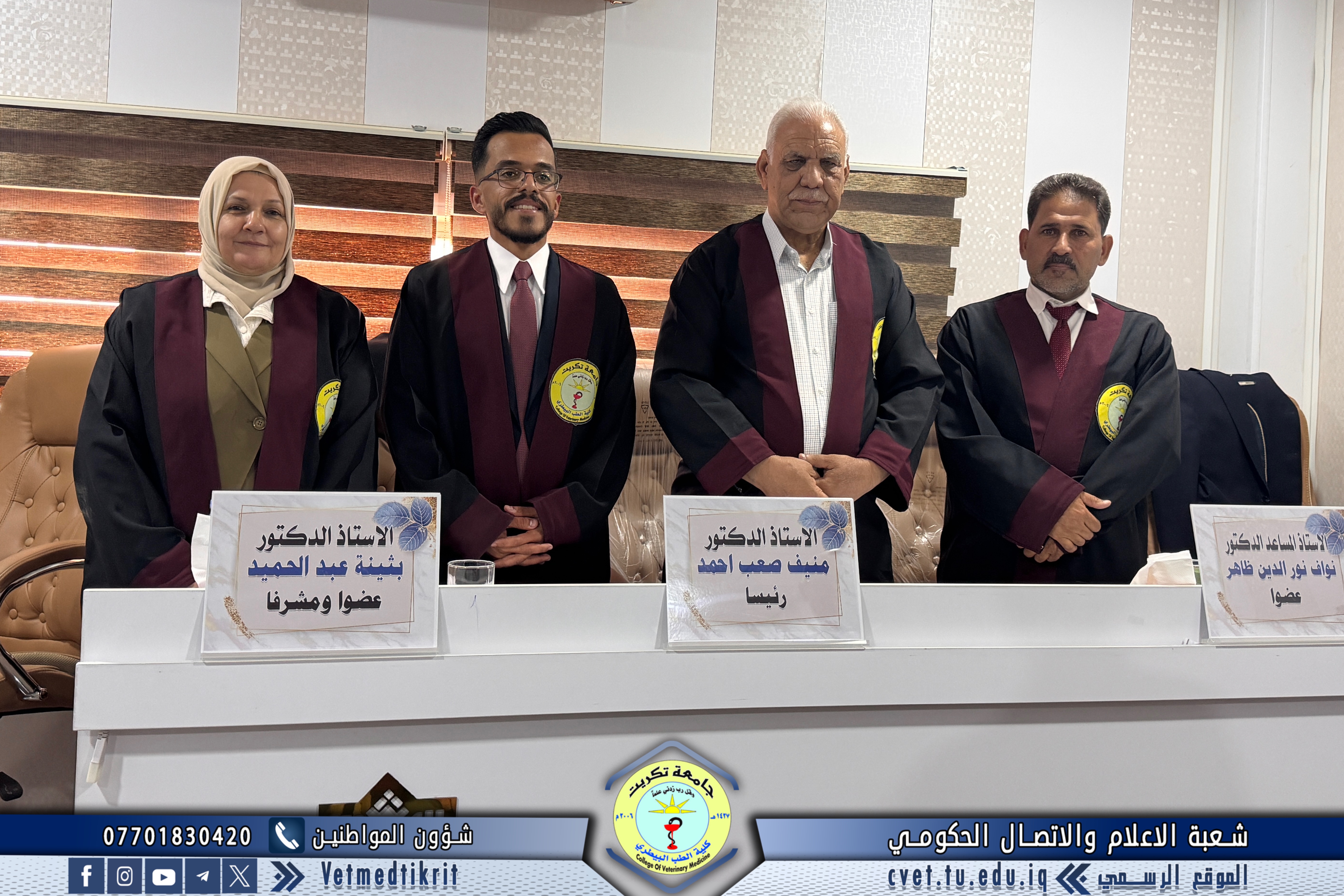
The examination committee consisted of:
- Prof. Dr. Iyad Hamid Ibrahim / Anatomy / University of Tikrit – College of Veterinary Medicine / Chair
- Prof. Dr. Kholoud Naji Rashid / Histology / University of Tikrit – College of Veterinary Medicine / Member
- Prof. Dr. Ammar Ghanim Mohammed / Anatomy and Histology / University of Mosul – College of Veterinary Medicine / Member
- Asst. Prof. Dr. Badr Khatlan Hamid / Anatomy and Histology / University of Tikrit – College of Veterinary Medicine / Member and Supervisor
Thesis Summary
The liver is one of the accessory organs of the digestive system, performing metabolic and secretory functions, including the secretion of bile, which is transported to the intestine through the biliary ducts. The gallbladder serves as a storage organ for bile, releasing it when needed.
The current study aimed to conduct an anatomical and histological comparison of the liver and the biliary duct system between two bird species—seagulls and pigeons—of both sexes and adult age groups. It also aimed to evaluate the branching patterns of the hepatic artery, hepatic vein, and portal vein within the livers of both species, as well as to assess the branching of the biliary duct network within the seagull’s liver.
A total of 70 birds (seagulls and pigeons, both sexes) were used. The samples were divided into four groups for each species:
Group One – Macroscopic Anatomy (20 birds)
Ten birds of each species (male and female) were used to study the gross anatomical structure of the liver, its measurements, and its relation to adjacent organs.
The results showed that the liver occupies a large portion of the thoracoabdominal cavity and is composed of two lobes (right and left). The right lobe was larger than the left in both species.
Group Two – Vascular Branching Study (20 birds)
Ten birds of each species were used to examine the branching of the hepatic artery, hepatic vein, and portal vein within the liver using latex injection technique.
In pigeons, the hepatic artery divides into two main branches (right and left) upon entering the liver.
In seagulls, the artery supplies the right lobe first, then connects to the left hepatic artery to supply the left lobe through a transverse segment.
Hepatic vein injection revealed:
In pigeons: three main branches (right, middle, and left).
In seagulls: two main branches (right and left) with a very small middle branch.
Portal vein injection in both species showed three main intrahepatic branches (right, transverse segment, and left), indicating similarity in the number of main branches but differences in the distribution of secondary branches.
Group Three – Biliary Duct System (10 birds)
Gross anatomical examination showed:
Seagulls possess a gallbladder with two bile ducts: the hepatointestinal duct and the cystointestinal duct.
Pigeons lack a gallbladder and the cystointestinal duct. Instead, they possess right and left hepatointestinal ducts.
Group Four – Histological Study (20 birds)
Ten birds of each species were examined using general and special stains.
The study found:
In both species, the liver is surrounded by Glisson’s capsule.
The liver parenchyma consists of irregularly shaped hepatocytes arranged in cords radiating from the central vein, forming clusters farther away from the center.
Sinusoids were wide in pigeons and narrow in seagulls.
Kupffer cells and lymphocytic aggregates were present, with variable distribution between the two species.
Binucleated hepatocytes were observed near the portal area in both species.
Ito cells were more numerous in seagulls than in pigeons.
Differences were noted in microscopic measurements of hepatic structures between both species.
In seagulls, intrahepatic bile ducts showed epithelial changes near the gallbladder.
The gallbladder epithelium was simple columnar.
Major bile ducts in pigeons and the cystointestinal duct in seagulls were lined with simple or stratified columnar epithelium.
The hepatointestinal duct in seagulls had simple cuboidal epithelium.
The study concluded that glycogen storage in the pigeon liver was higher than in the seagull liver, while connective tissue fibers in the portal area were more abundant in seagulls.
The defense session, held in Dr. Muhannad Maher’s Hall at the College of Veterinary Medicine, was attended by several faculty members and a number of students.