


By the grace of God, the Master's thesis entitled "Histological Changes Associated with Exposure to Lead Acetate and Evaluation of the Therapeutic Role of Berbin Extract for Female Albino Rats and Their Fetuses" was discussed by the student (Ayad Salem Mahdi Saleh) specializing in (Veterinary Histology and Embryology) at the College of Veterinary Medicine at Tikrit University, in the presence of Professor Dr. Bashar Sadiq Noumi, Dean of the College. The discussion committee consisted of:
- Professor Dr. Ayad Hamid Ibrahim, Anatomy Department, Tikrit University, College of Veterinary Medicine, Chairman
- Assistant Professor Dr. Badr Khatblan Hamid, Anatomy and Histology Department, Tikrit University, College of Veterinary Medicine, Member
- Assistant Professor Dr. Rashid Khamis Shaaban, Histology and Embryology Department, Tikrit University, College of Education for Pure Sciences, Member
- Assistant Professor Dr. Mahmoud Nawfal Mustafa, Histology and Embryology Department, Tikrit University, College of Veterinary Medicine, Member and Supervisor
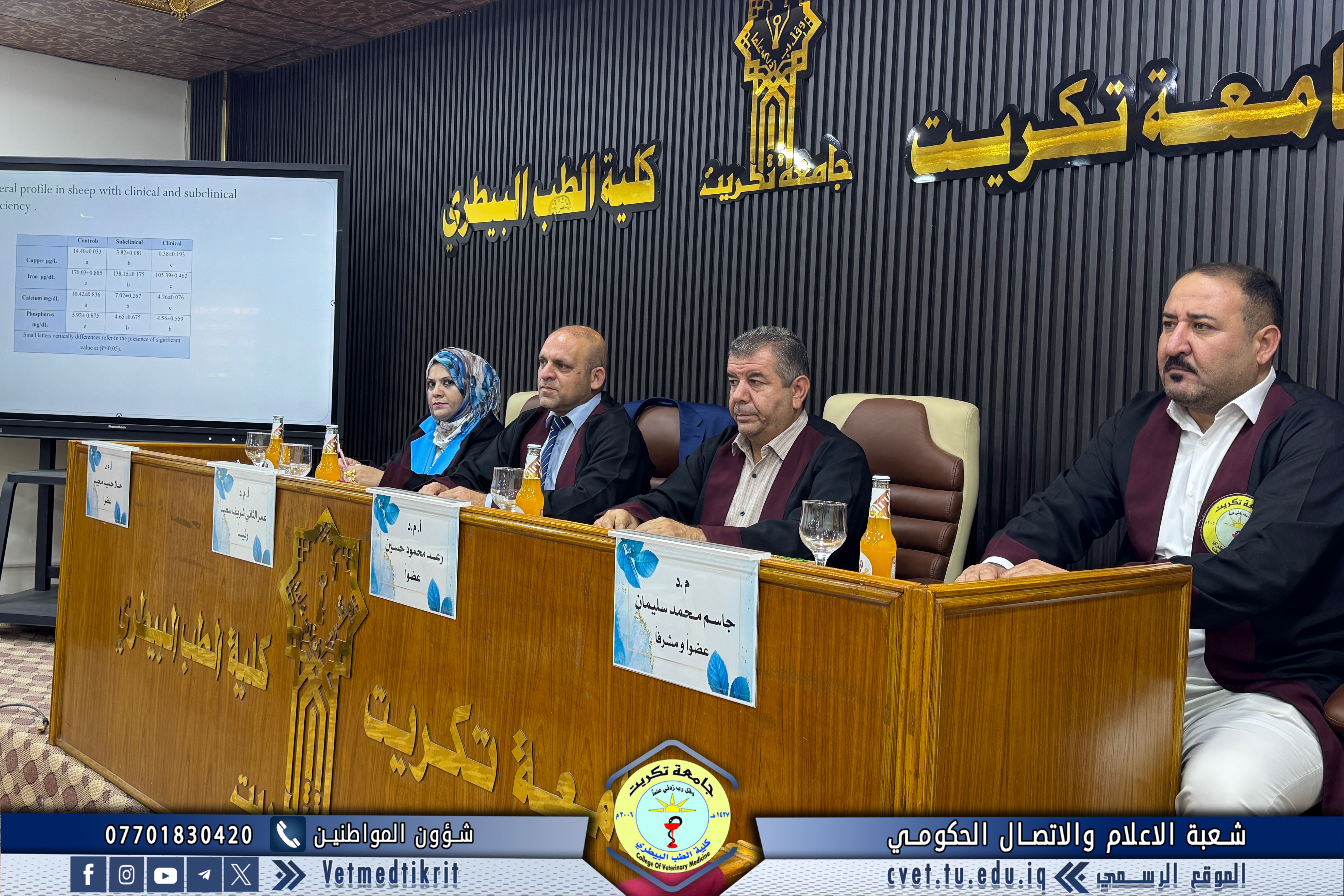
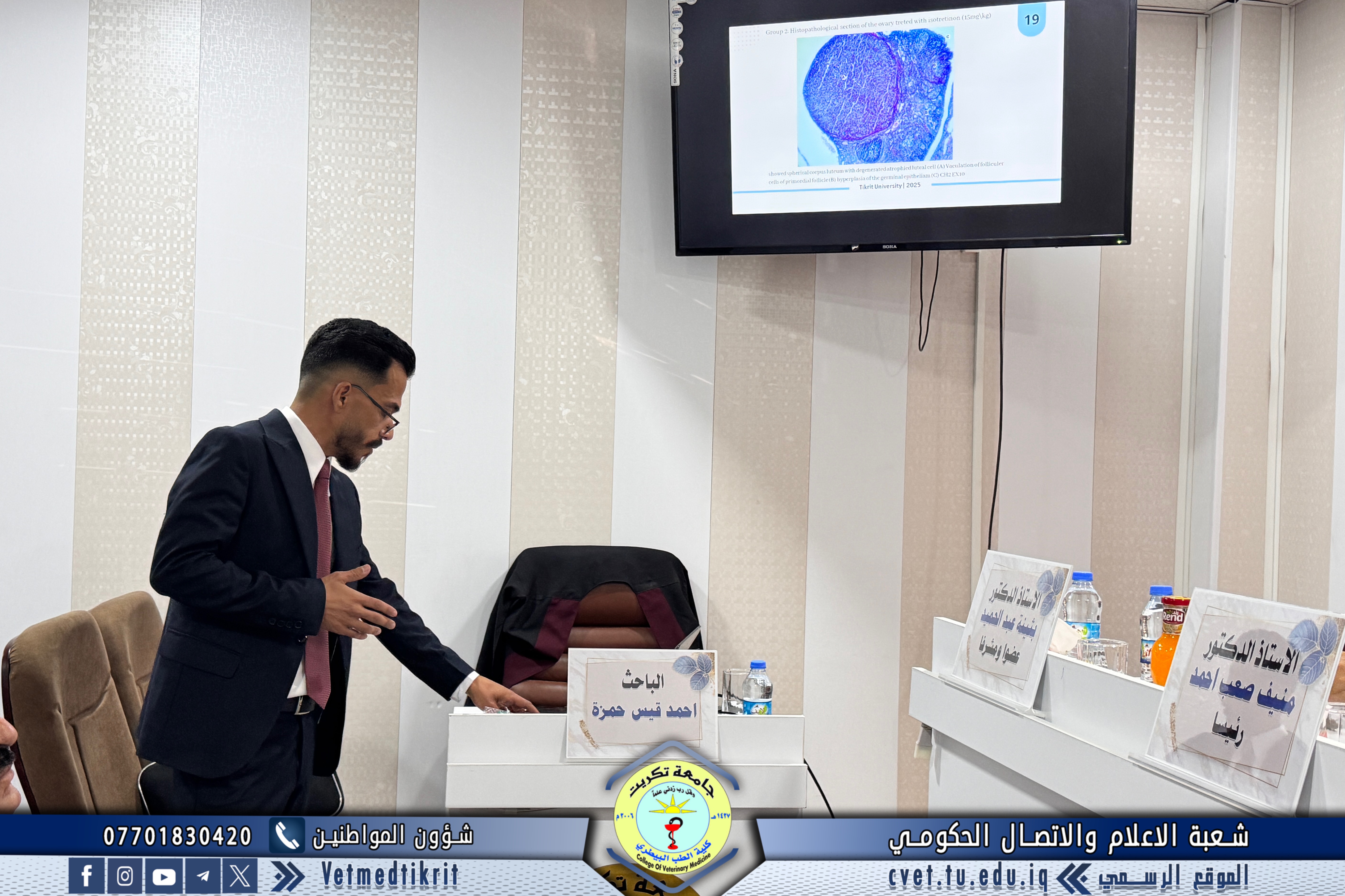
The summary of the thesis was as follows: The current study was conducted in the animal house of the College of Veterinary Medicine at Tikrit University, from October 27, 2024, to November 27, 2024. The study included an investigation into the effect of lead acetate on inducing histological changes in pregnant female albino rats and their fetuses, and its therapeutic role. For the aqueous extract of purslane plant. (30) female white rats, aged between 10-12 weeks and weighing between 180-200 grams, were used. The females were placed in plastic cages with males of the same strain for the purpose of mating. The vaginal plug was examined to ensure that mating had occurred, as this examination is evidence of fertilization. The day of mating was considered day zero of pregnancy. The animals were then randomly distributed into six groups that were treated as follows: the first group (control group) was treated with distilled water, the second group (negative control) was treated with lead acetate during the period 4-9 days of pregnancy at a dose of 50 mg/kg, the third group (negative control) was treated with lead acetate during the period 10-19 days of pregnancy at a dose of 50 mg/kg, the fourth group (positive control) was treated with the aqueous extract of the berbin plant throughout the pregnancy period at a dose of 200 mg/kg, the fifth group was treated with the aqueous extract of the berbin plant throughout the pregnancy period at a dose of 200 mg/kg and was also treated with lead acetate during the period 4-9 days of pregnancy at a dose of 50 mg/kg, and the sixth group was treated with the aqueous extract of the berbin plant throughout the pregnancy period at a dose of 200 mg/kg and was also treated with lead acetate during the period 10-19 days of gestation at a dose of 50 mg/kg, administered orally once daily by gavage. The experimental animals were fed a standard diet. On day 20 of gestation, the animals were dissected, and the liver and kidneys of each animal were extracted. Blood was withdrawn and embryos were extracted for histological and physiological examinations and morphological assessment of each embryo in the studied groups. Liver changes were manifested by cytoplasmic vacuolar degeneration with hypertrophic hepatocytes, dilated blood sinusoids, hypervolemia of Koffer cells, infiltration of inflammatory cells, hepatocellular atrophy and hematoma. Renal histological changes included glomerular atrophy with leukocyte infiltration, dilated capsular space, glomerular disappearance, convoluted tubule cell destruction, hemorrhage within the renal tissue, irregular tubular size, congestion, glomerular atrophy, convoluted tubule degeneration, and glomerular atrophy. Physiological tests in the current study also showed a significant decrease in serum superoxide dismutase concentrations in groups G2 and G3, no decrease in group G4, and a slight significant decrease in groups G5 and G6. These readings returned to near-normal levels when compared with the control group G1. The results of the current study showed a significant increase in the concentration of malondialdehyde in groups G2 and G3 treated with lead acetate only, and a significant decrease in group G4 treated with the aqueous extract of berbene plant only, and no significant difference was observed between groups G5 and G6 treated with lead acetate and berbene plant aqueous extract compared with the control group G1. The results of the current study also showed a significant increase in the concentration of gamma-glutamyl transferase in the blood serum of both groups G2 and G3, and no significant differences were observed between group G4 treated with the aqueous extract of berbene plant and groups G5 and G6 treated with lead acetate and berbene plant aqueous extract compared with the control group. The results of the current study also showed a significant increase in serum creatinine concentrations in groups G2 and G3 treated with lead acetate, and a significant decrease in group G4 treated with aqueous berberine extract, and groups G5 and G6 treated with lead acetate and aqueous berberine extract, compared to the control group G1. The results of the morphological changes in the embryos showed short fetuses, trunk and tail torsion with internal congestion, short limbs, a short snout, and skin wrinkles in groups G2 and G3 exposed to lead acetate over two different periods. The fetuses in group G4 treated with only aqueous berberine extract were normal, while the fetuses in groups G5 and G6 exposed to lead acetate over two different periods, as well as aqueous berberine extract, were nearly normal, with slight trunk and tail torsion and short stature, compared to the control group G1.
The discussion, which was held in Dr. Muhannad Maher Hall at the College of Veterinary Medicine, was attended by a number of female and male faculty members and a group of students.